Papilledema physical examination
|
Papilledema |
|
Diagnosis |
|---|
|
Treatment |
|
Case Studies |
|
Papilledema physical examination On the Web |
|
American Roentgen Ray Society Images of Papilledema physical examination |
|
Risk calculators and risk factors for Papilledema physical examination |
Editor-In-Chief: C. Michael Gibson, M.S., M.D. [1] Associate Editor(s)-In-Chief:Kalsang Dolma, MBBS
overview
Physical Examination
Eyes
-
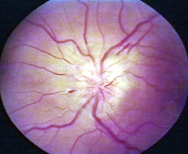
Papilledema.
-
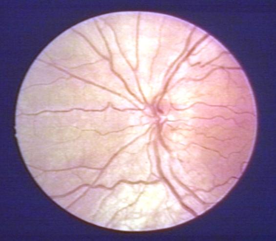
Pseudopapilledema.


There are 10 hallmarks of papilledema:
- Blurring of the disc margins
- Filling in of the optic disc cup
- Anterior bulging of the nerve head
- Edema of the nerve fiber layer
- Retinal or choroidal folds
- Congestion of retinal veins
- Peripapillary hemorrhages
- Hyperemia of the optic nerve head
- Nerve fiber layer infarcts
- Hard exudates of the optic disc