Non small cell lung cancer pathophysiology: Difference between revisions
No edit summary |
|||
| Line 9: | Line 9: | ||
*The pathogenesis of non-small cell lung cancer will depend on the type of histological subtype of lung cancer: lung adenocarcinoma, lung large cell carcinoma, and squamous cell lung carcinoma.<ref name="pmid16107574">{{cite journal |vauthors=Miller YE |title=Pathogenesis of lung cancer: 100 year report |journal=Am. J. Respir. Cell Mol. Biol. |volume=33 |issue=3 |pages=216–23 |year=2005 |pmid=16107574 |pmc=2715312 |doi=10.1165/rcmb.2005-0158OE |url=}}</ref> | *The pathogenesis of non-small cell lung cancer will depend on the type of histological subtype of lung cancer: lung adenocarcinoma, lung large cell carcinoma, and squamous cell lung carcinoma.<ref name="pmid16107574">{{cite journal |vauthors=Miller YE |title=Pathogenesis of lung cancer: 100 year report |journal=Am. J. Respir. Cell Mol. Biol. |volume=33 |issue=3 |pages=216–23 |year=2005 |pmid=16107574 |pmc=2715312 |doi=10.1165/rcmb.2005-0158OE |url=}}</ref> | ||
*Non-small cell lung cancer arises from the [[epithelial]] cells of the lung of the central [[bronchi]] to terminal [[alveoli]], which are normally involved in the protection of the airways. | *Non-small cell lung cancer arises from the [[epithelial]] cells of the lung of the central [[bronchi]] to terminal [[alveoli]], which are normally involved in the protection of the airways. | ||
*Lung adenocarcinoma arises from the bronchial mucosal glands of the lung (main, lobar, segmental bronchi, or lung parenchyma), which are normally involved in decreasing bacterial growth in the epithelial cells of the lung. | *Lung adenocarcinoma arises from the bronchial mucosal glands of the lung (main, lobar, [[Bronchi|segmental bronchi]], or [[Parenchyma|lung parenchyma]]), which are normally involved in decreasing bacterial growth in the epithelial cells of the lung. | ||
*Lung adenocarcinoma usually occurs in a peripheral location within the lung. In the majority of the patients, cancer arises from precursor lesions, such as pre-existing scars or inflammation (chronic infections). | *Lung adenocarcinoma usually occurs in a peripheral location within the lung. In the majority of the patients, cancer arises from precursor lesions, such as pre-existing scars or inflammation (chronic infections). | ||
*Lung adenocarcinoma can also result from multiple genetic mutations. For more information about lung adenocarcinoma pathogenesis, see [[Adenocarcinoma of the lung pathophysiology|here]] | *Lung adenocarcinoma can also result from multiple genetic mutations. For more information about lung adenocarcinoma pathogenesis, see [[Adenocarcinoma of the lung pathophysiology|here]] | ||
*Squamous lung cell carcinoma arises from bronchial epithelial cell damage (related with active smoking). Squamous cell carcinoma usually occurs in a central location within the lung. For more information about squamous lung cell carcinoma pathogenesis, see [[Squamous cell carcinoma of the lung pathophysiology|here]] | *Squamous lung cell carcinoma arises from bronchial epithelial cell damage (related with active smoking). Squamous cell carcinoma usually occurs in a central location within the lung. For more information about squamous lung cell carcinoma pathogenesis, see [[Squamous cell carcinoma of the lung pathophysiology|here]] | ||
*Large cell carcinoma of the lung arises from large polygonal and anaplastic cells, hard to differentiate from other types of non small cell lung cancer. Large cell carcinoma usually occurs in a peripheral location within the lung. | *Large cell carcinoma of the lung arises from large polygonal and anaplastic cells, hard to differentiate from other types of non small cell lung cancer. Large cell carcinoma usually occurs in a peripheral location within the lung. | ||
*Non-small cell lung cancer is an invasive and rapidly growing cancer which may metastasize to different organs of the body. | *Non-small cell lung cancer is an invasive and rapidly growing cancer which may [[Metastasis|metastasize]] to different organs of the body. | ||
==Genetics== | ==Genetics== | ||
| Line 21: | Line 21: | ||
* The table below describes the [[gene]]s involved in the pathogenesis of non small cell lung cancer. | * The table below describes the [[gene]]s involved in the pathogenesis of non small cell lung cancer. | ||
{| style="border: 0px; font-size: 90%; margin: 3px; width: 800px" align=center | {| style="border: 0px; font-size: 90%; margin: 3px; width: 800px" align="center" | ||
|valign=top| | | valign="top" | | ||
|+ | |+ | ||
!style="background: #4479BA; width: 20px;"|{{fontcolor|#FFF|'''Genes'''}} | ! style="background: #4479BA; width: 20px;" |{{fontcolor|#FFF|'''Genes'''}} | ||
!style="background: #4479BA; width: 100px;"|{{fontcolor|#FFF|'''Presence in non small cell-lung cancers'''}} | ! style="background: #4479BA; width: 100px;" |{{fontcolor|#FFF|'''Presence in non small cell-lung cancers'''}} | ||
|- | |- | ||
|style="padding: 0 5px; background: #DCDCDC" align=center"|'''[[EGFR]]''' | | style="padding: 0 5px; background: #DCDCDC" align="center"" |'''[[EGFR]]''' | ||
|style="padding: 5px 5px; background: #F5F5F5;"| | | style="padding: 5px 5px; background: #F5F5F5;" | | ||
*EGFR mutations are present in approximately 10% to 15% of all non-small cell lung cancers | *EGFR mutations are present in approximately 10% to 15% of all non-small cell lung cancers | ||
|- | |- | ||
| style="padding: 0 5px; background: #DCDCDC"| '''[[KRAS]]''' | | style="padding: 0 5px; background: #DCDCDC" | '''[[KRAS]]''' | ||
| style="padding: 5px 5px; background: #F5F5F5;"| | | style="padding: 5px 5px; background: #F5F5F5;" | | ||
* Mutations are present in approximately 30% of pulmonary adenocarcinomas | * Mutations are present in approximately 30% of pulmonary adenocarcinomas | ||
*Mutations are present in approximately 5% of pulmonary squamous cell carcinomas | *Mutations are present in approximately 5% of pulmonary squamous cell carcinomas | ||
*Associated with carcinomas with mucinous histology | *Associated with carcinomas with mucinous histology | ||
|- | |- | ||
| style="padding: 0 5px; background: #DCDCDC" |'''ALK''' | | style="padding: 0 5px; background: #DCDCDC" |'''[[ALK-negative ALCL|ALK]]''' | ||
|style="padding: 5px 5px; background: #F5F5F5;"| | | style="padding: 5px 5px; background: #F5F5F5;" | | ||
*Mutations are present in approximately 5% of all non-small cell lung cancers | *Mutations are present in approximately 5% of all non-small cell lung cancers | ||
|- | |- | ||
|style="padding: 0 5px; background: #DCDCDC" | '''[[HER2]]''' | | style="padding: 0 5px; background: #DCDCDC" | '''[[HER2]]''' | ||
|style="padding: 5px 5px; background: #F5F5F5;"| | | style="padding: 5px 5px; background: #F5F5F5;" | | ||
*Mutations are present in approximately 4% of adenocarcinomas | *Mutations are present in approximately 4% of adenocarcinomas | ||
|- | |- | ||
| style="padding: 0 5px; background: #DCDCDC" |'''[[BRAF]]''' | | style="padding: 0 5px; background: #DCDCDC" |'''[[BRAF]]''' | ||
| style="padding: 5px 5px; background: #F5F5F5;"| | | style="padding: 5px 5px; background: #F5F5F5;" | | ||
*Mutations are present in less than 2% of adenocarcinomas | *Mutations are present in less than 2% of adenocarcinomas | ||
|- | |- | ||
| style="padding: 0 5px; background: #DCDCDC" |'''ROS-1''' | | style="padding: 0 5px; background: #DCDCDC" |'''[[ROS1 (gene)|ROS-1]]''' | ||
| style="padding: 5px 5px; background: #F5F5F5;"| | | style="padding: 5px 5px; background: #F5F5F5;" | | ||
*Mutations are present in less than 2% of adenocarcinomas | *Mutations are present in less than 2% of adenocarcinomas | ||
|} | |} | ||
| Line 57: | Line 57: | ||
*Other conditions associated with non-small cell lung cancer, include: | *Other conditions associated with non-small cell lung cancer, include: | ||
*Genetic conditions | *Genetic conditions | ||
:* | :*Li–Fraumeni syndrome | ||
:*[[Noonan syndrome]] | :*[[Noonan syndrome]] | ||
*Infections | *Infections | ||
:*Human papillomavirus (HPV) infection | :*[[Human papillomavirus|Human papillomavirus (HPV) infection]] | ||
:*[[Pulmonary tuberculosis]] | :*[[Pulmonary tuberculosis]] | ||
| Line 66: | Line 66: | ||
*On gross pathology, findings will depend on the histological subtype of non-small cell lung cancer. | *On gross pathology, findings will depend on the histological subtype of non-small cell lung cancer. | ||
*Lung adenocarcinoma gross pathology findings, include:<ref name="pathology"> Non small cell lung cancer. Libre Pathology. http://librepathology.org/wiki/Non-small_cell_lung_carcinoma Accessed on February 22, 2016 </ref> | *Lung adenocarcinoma gross pathology findings, include:<ref name="pathology">Non small cell lung cancer. Libre Pathology. http://librepathology.org/wiki/Non-small_cell_lung_carcinoma Accessed on February 22, 2016 </ref> | ||
:*Spherical tumor with well-defined borders | :*Spherical tumor with well-defined borders | ||
:*Homogeneous gray-white cut surface | :*Homogeneous gray-white cut surface | ||
:*Involvement of the thoracic wall | :*Involvement of the [[thoracic wall]] | ||
:*Usually found in the peripheral lung | :*Usually found in the peripheral lung | ||
*Large cell lung cancer gross pathology findings, include:<ref name="pathology"> Non small cell lung cancer. Libre Pathology. http://librepathology.org/wiki/Non-small_cell_lung_carcinoma Accessed on February 22, 2016 </ref> | *Large cell lung cancer gross pathology findings, include:<ref name="pathology">Non small cell lung cancer. Libre Pathology. http://librepathology.org/wiki/Non-small_cell_lung_carcinoma Accessed on February 22, 2016 </ref> | ||
:*Well-defined borders | :*Well-defined borders | ||
:*Resemblance to gross findings in adenocarcinoma | :*Resemblance to gross findings in adenocarcinoma | ||
:*No signs of anthracosis | :*No signs of [[anthracosis]] | ||
:*Involvement of the thoracic wall | :*Involvement of the thoracic wall | ||
*Squamous cell lung cancer gross pathology findings, include:<ref name="pathology"> Non small cell lung cancer. Libre Pathology. http://librepathology.org/wiki/Non-small_cell_lung_carcinoma Accessed on February 22, 2016 </ref> | *Squamous cell lung cancer gross pathology findings, include:<ref name="pathology">Non small cell lung cancer. Libre Pathology. http://librepathology.org/wiki/Non-small_cell_lung_carcinoma Accessed on February 22, 2016 </ref> | ||
:*Lung mass | :*Lung mass | ||
:*Usually centrally located | :*Usually centrally located | ||
:*Associated with a large airway | :*Associated with a large airway | ||
:*Usually have a central cavitation | :*Usually have a [[Cavitation|central cavitation]] | ||
==Microscopic Pathology== | ==Microscopic Pathology== | ||
| Line 86: | Line 86: | ||
*On microscopic pathology, findings will depend on the histological type of non-small cell lung cancer. | *On microscopic pathology, findings will depend on the histological type of non-small cell lung cancer. | ||
*Lung adenocarcinoma microscopic pathology findings, include:<ref name="pathology"> Non small cell lung cancer. Libre Pathology. http://librepathology.org/wiki/Non-small_cell_lung_carcinoma Accessed on February 22, 2016 </ref> | *Lung adenocarcinoma microscopic pathology findings, include:<ref name="pathology">Non small cell lung cancer. Libre Pathology. http://librepathology.org/wiki/Non-small_cell_lung_carcinoma Accessed on February 22, 2016 </ref> | ||
:*Nuclear atypia | :*Nuclear atypia | ||
:*Eccentrically placed nuclei | :*Eccentrically placed nuclei | ||
| Line 94: | Line 94: | ||
:*Different patterns, include: acinar, lepidic, micropapillary, papillary, and solid. | :*Different patterns, include: acinar, lepidic, micropapillary, papillary, and solid. | ||
*Large cell lung cancer microscopic pathology findings, include:<ref name="pathology"> Non small cell lung cancer. Libre Pathology. http://librepathology.org/wiki/Non-small_cell_lung_carcinoma Accessed on February 22, 2016 </ref> | *Large cell lung cancer microscopic pathology findings, include:<ref name="pathology">Non small cell lung cancer. Libre Pathology. http://librepathology.org/wiki/Non-small_cell_lung_carcinoma Accessed on February 22, 2016 </ref> | ||
:*Large polygonal cells and anaplastic cells | :*Large polygonal cells and anaplastic cells | ||
:*No squamous or glandular differentiation | :*No squamous or glandular differentiation | ||
| Line 100: | Line 100: | ||
:*Vesicular nuclei, prominent nucleoli | :*Vesicular nuclei, prominent nucleoli | ||
*Squamous cell lung cancer microscopic pathology findings, include:<ref name="pathology"> Non small cell lung cancer. Libre Pathology. http://librepathology.org/wiki/Non-small_cell_lung_carcinoma Accessed on February 22, 2016 </ref> | *Squamous cell lung cancer microscopic pathology findings, include:<ref name="pathology">Non small cell lung cancer. Libre Pathology. http://librepathology.org/wiki/Non-small_cell_lung_carcinoma Accessed on February 22, 2016 </ref> | ||
:*Central nucleus | :*Central nucleus | ||
:*Dense appearing cytoplasm, usually eosinophilic | :*Dense appearing cytoplasm, usually eosinophilic | ||
| Line 107: | Line 107: | ||
*On inmunohistochemistry, findings will depend on the histological type of non-small cell lung cancer. | *On inmunohistochemistry, findings will depend on the histological type of non-small cell lung cancer. | ||
*Common inmunohistochemistry markers used for non-small cell carcinoma subtyping, include:<ref name="pathology"> Non small cell lung cancer. Libre Pathology. http://librepathology.org/wiki/Non-small_cell_lung_carcinoma Accessed on February 22, 2016 </ref> | *Common inmunohistochemistry markers used for non-small cell carcinoma subtyping, include:<ref name="pathology">Non small cell lung cancer. Libre Pathology. http://librepathology.org/wiki/Non-small_cell_lung_carcinoma Accessed on February 22, 2016 </ref> | ||
:*TTF-1 for adenocarcinoma | :*TTF-1 for adenocarcinoma | ||
:*p63 and high-molecular weight keratins for squamous cell carcinoma | :*p63 and high-molecular weight keratins for squamous cell carcinoma | ||
:*Lack of staining with neuroendocrine markers (chromogranin A, synaptophysin, and CD56) | :*Lack of staining with neuroendocrine markers ([[chromogranin A]], synaptophysin, and [[CD56]]) | ||
==Gallery== | ==Gallery== | ||
| Line 127: | Line 127: | ||
==References== | ==References== | ||
{{reflist|2}} | {{reflist|2}} | ||
[[Category:Types of cancer]] | [[Category:Types of cancer]] | ||
Revision as of 17:00, 23 February 2016
|
Non Small Cell Lung Cancer Microchapters |
|
Differentiating Non Small Cell Lung Cancer from other Diseases |
|---|
|
Diagnosis |
|
Treatment |
|
Case Studies |
|
Non small cell lung cancer pathophysiology On the Web |
|
American Roentgen Ray Society Images of Non small cell lung cancer pathophysiology |
|
Directions to Hospitals Treating Non small cell carcinoma of the lung |
|
Risk calculators and risk factors for Non small cell lung cancer pathophysiology |
Editor-In-Chief: C. Michael Gibson, M.S., M.D. [1] Associate Editor(s)-in-Chief: Maria Fernanda Villarreal, M.D. [2]
Overview
Non-small cell lung cancer arises from the epithelial cells of the lung of the central bronchi to terminal alveoli, which are normally involved in the protection of the airways. Non-small cell lung cancer is an invasive and rapidly growing cancer which may metastasize to different organs of the body. Genes involved in the pathogenesis of non-small cell lung cancer include EGFR, KRAS, HER2, BRAF, and ALK. On gross pathology, findings will depend on the histological type. On microscopic histopathological analysis non-small cell lung cancer demonstrate large cells with abundant cytoplasm and no stippled chromatin.[1]
Pathogenesis
- The pathogenesis of non-small cell lung cancer will depend on the type of histological subtype of lung cancer: lung adenocarcinoma, lung large cell carcinoma, and squamous cell lung carcinoma.[1]
- Non-small cell lung cancer arises from the epithelial cells of the lung of the central bronchi to terminal alveoli, which are normally involved in the protection of the airways.
- Lung adenocarcinoma arises from the bronchial mucosal glands of the lung (main, lobar, segmental bronchi, or lung parenchyma), which are normally involved in decreasing bacterial growth in the epithelial cells of the lung.
- Lung adenocarcinoma usually occurs in a peripheral location within the lung. In the majority of the patients, cancer arises from precursor lesions, such as pre-existing scars or inflammation (chronic infections).
- Lung adenocarcinoma can also result from multiple genetic mutations. For more information about lung adenocarcinoma pathogenesis, see here
- Squamous lung cell carcinoma arises from bronchial epithelial cell damage (related with active smoking). Squamous cell carcinoma usually occurs in a central location within the lung. For more information about squamous lung cell carcinoma pathogenesis, see here
- Large cell carcinoma of the lung arises from large polygonal and anaplastic cells, hard to differentiate from other types of non small cell lung cancer. Large cell carcinoma usually occurs in a peripheral location within the lung.
- Non-small cell lung cancer is an invasive and rapidly growing cancer which may metastasize to different organs of the body.
Genetics
- Development of non-small cell lung cancer is the result of multiple genetic mutations.
- Genetic mutations play an important role in the treatment selection for non small cell lung cancer.
- The table below describes the genes involved in the pathogenesis of non small cell lung cancer.
| Genes | Presence in non small cell-lung cancers |
|---|---|
| EGFR |
|
| KRAS |
|
| ALK |
|
| HER2 |
|
| BRAF |
|
| ROS-1 |
|
Associated Conditions
- Other conditions associated with non-small cell lung cancer, include:
- Genetic conditions
- Li–Fraumeni syndrome
- Noonan syndrome
- Infections
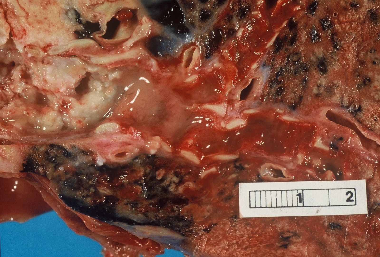
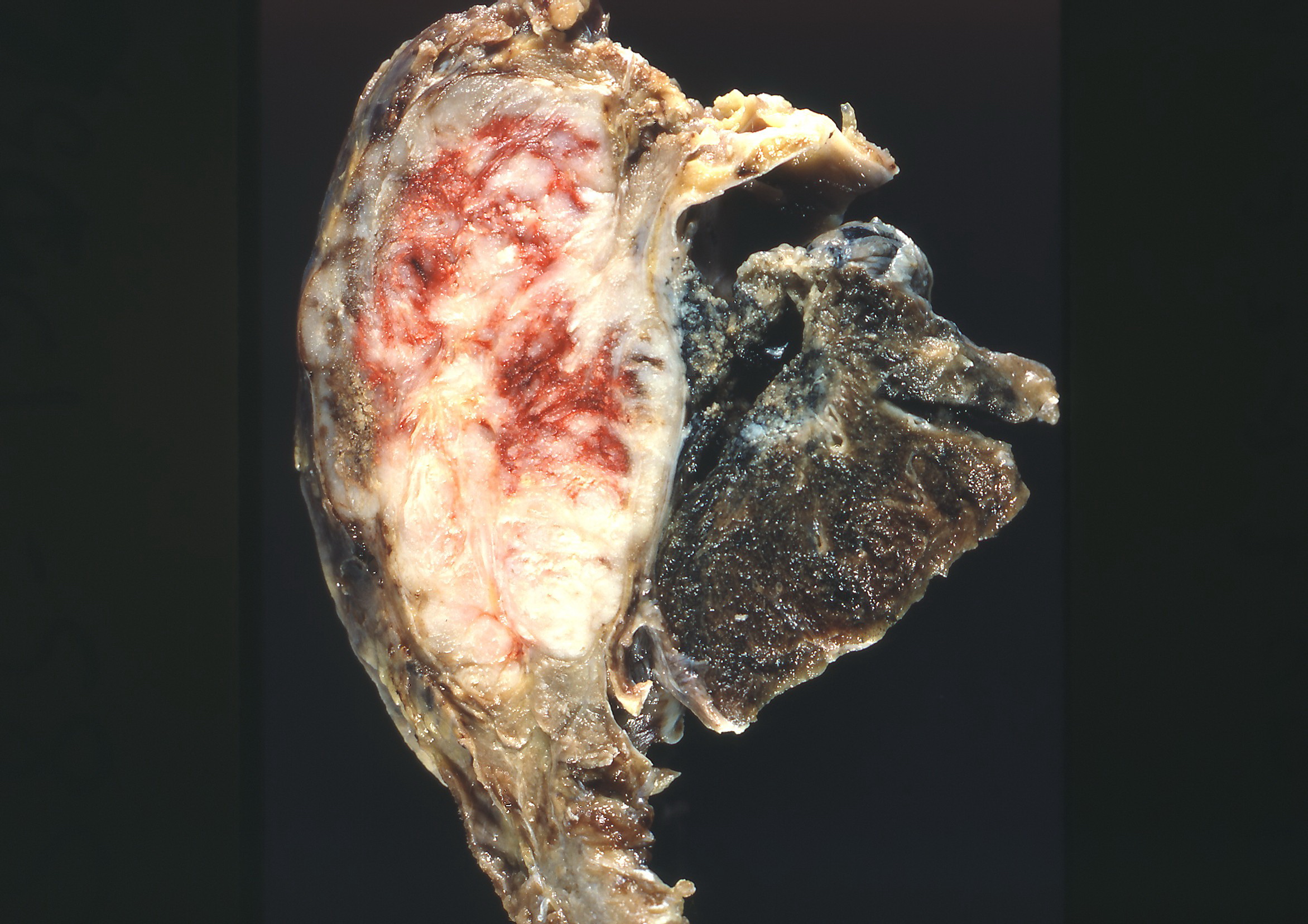
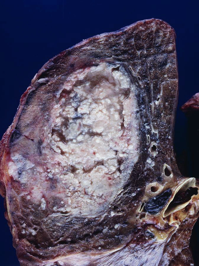
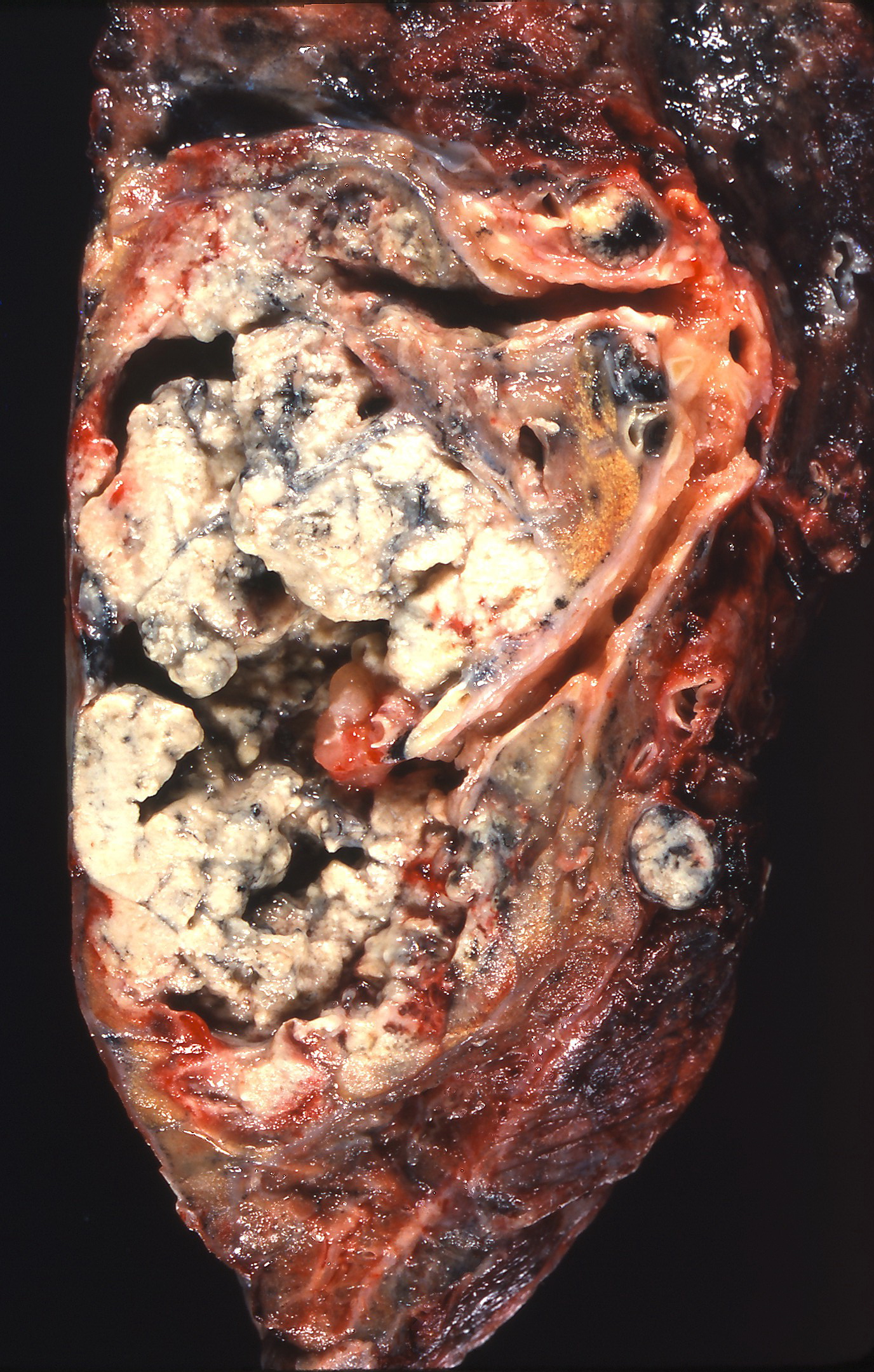
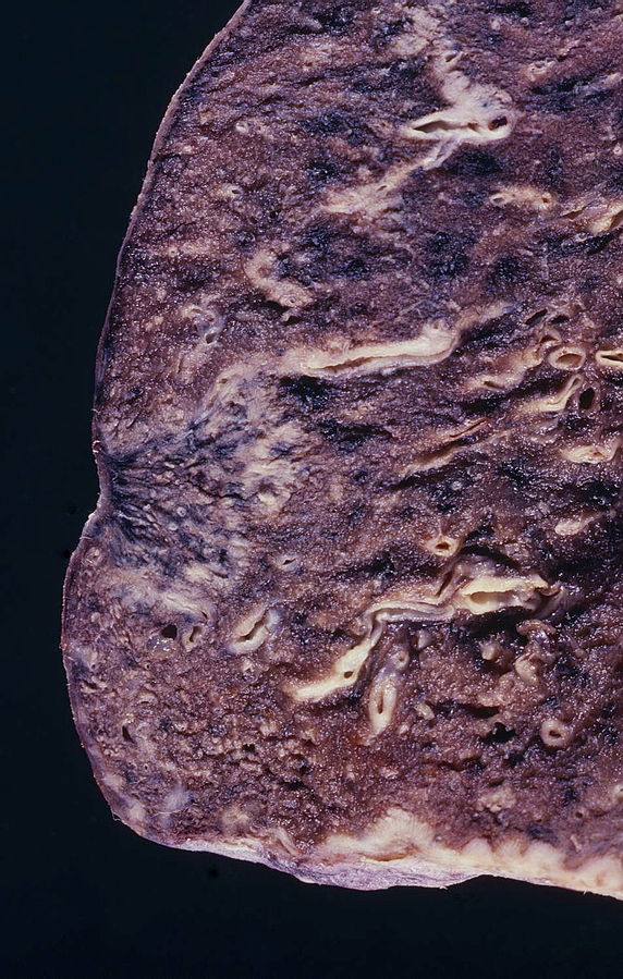
Gross Pathology
- On gross pathology, findings will depend on the histological subtype of non-small cell lung cancer.
- Lung adenocarcinoma gross pathology findings, include:[2]
- Spherical tumor with well-defined borders
- Homogeneous gray-white cut surface
- Involvement of the thoracic wall
- Usually found in the peripheral lung
- Large cell lung cancer gross pathology findings, include:[2]
- Well-defined borders
- Resemblance to gross findings in adenocarcinoma
- No signs of anthracosis
- Involvement of the thoracic wall
- Squamous cell lung cancer gross pathology findings, include:[2]
- Lung mass
- Usually centrally located
- Associated with a large airway
- Usually have a central cavitation
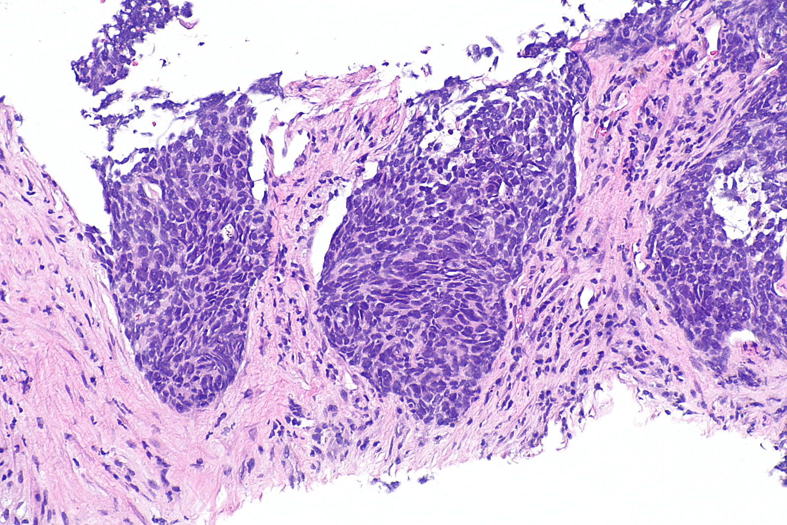
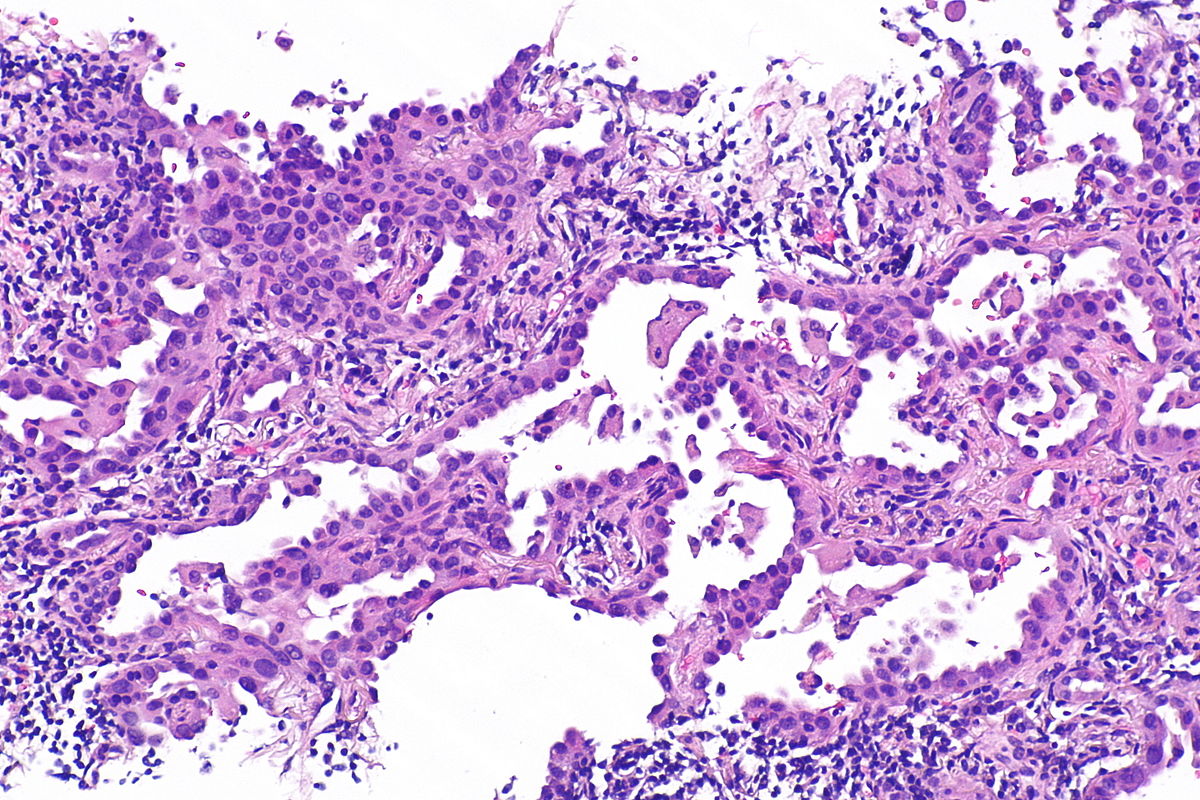
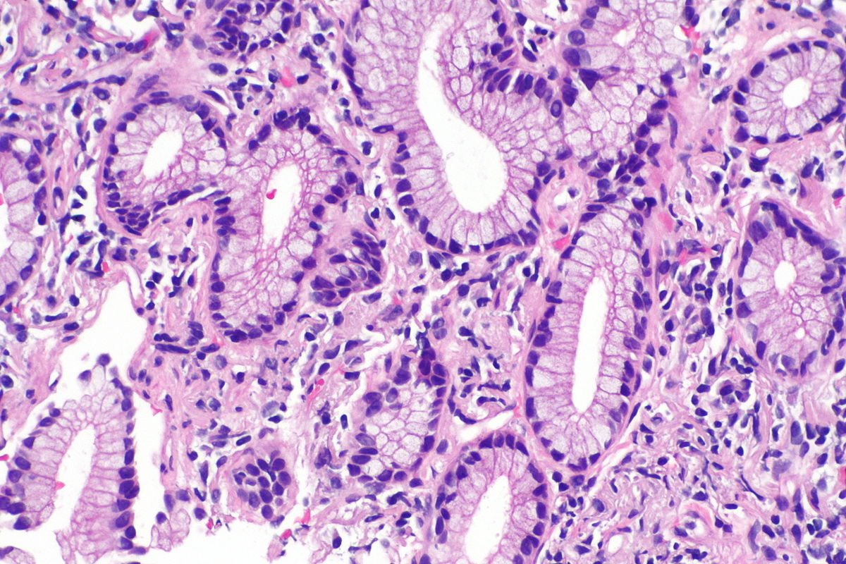
Microscopic Pathology
- On microscopic pathology, findings will depend on the histological type of non-small cell lung cancer.
- Lung adenocarcinoma microscopic pathology findings, include:[2]
- Nuclear atypia
- Eccentrically placed nuclei
- Abundant cytoplasm with mucin vacuoles
- Often conspicuous nucleoli
- Lack of intercellular bridges.
- Different patterns, include: acinar, lepidic, micropapillary, papillary, and solid.
- Large cell lung cancer microscopic pathology findings, include:[2]
- Large polygonal cells and anaplastic cells
- No squamous or glandular differentiation
- Moderately abundant cytoplasm
- Vesicular nuclei, prominent nucleoli
- Squamous cell lung cancer microscopic pathology findings, include:[2]
- Central nucleus
- Dense appearing cytoplasm, usually eosinophilic
- Small nucleolus
- Intracellular bridges (classic feature)
- On inmunohistochemistry, findings will depend on the histological type of non-small cell lung cancer.
- Common inmunohistochemistry markers used for non-small cell carcinoma subtyping, include:[2]
- TTF-1 for adenocarcinoma
- p63 and high-molecular weight keratins for squamous cell carcinoma
- Lack of staining with neuroendocrine markers (chromogranin A, synaptophysin, and CD56)
Gallery
-
Gross pathology:Bronchial squamous lung cell cancer
-
Gross pathology: adenocarcinoma of the lung
-
Gross pathology: Squamous lung cell cancer
-
Gross pathology: cavitation, squamous lung cell cancer
-
Gross pathology: adenocarcinoma of the lung
-
Micropathology non-small cell lung cancer
-
Micrograph of mucinous adenocarcinoma of the lung. H&E stain.
-
Micrograph showing an adenocarcinoma of the lung (acinar pattern). H&E stain.


References
- ↑ 1.0 1.1 Miller YE (2005). "Pathogenesis of lung cancer: 100 year report". Am. J. Respir. Cell Mol. Biol. 33 (3): 216–23. doi:10.1165/rcmb.2005-0158OE. PMC 2715312. PMID 16107574.
- ↑ 2.0 2.1 2.2 2.3 2.4 2.5 2.6 Non small cell lung cancer. Libre Pathology. http://librepathology.org/wiki/Non-small_cell_lung_carcinoma Accessed on February 22, 2016