Mitral stenosis electrocardiogram: Difference between revisions

Jump to navigation Jump to search
No edit summary
No edit summary
Line 2: Line 2:
{{CMG}}
{{CMG}}


'''Associate Editor-In-Chief:''' {{CZ}}
'''Associate Editor-In-Chief:''' {{CZ}}; [[Varun Kumar]], M.B.B.S.; [[Lakshmi Gopalakrishnan]], M.B.B.S.


{{Editor Help}}
{{Editor Help}}


===Electrocardiographic Findings===
===Electrocardiographic Findings in [[Mitral stenosis]]===
*[[LA enlargement]] ([[P mitrale]]): Left atrial enlargement produces a broad, bifid P wave in lead II (P mitrale) and enlarges the terminal negative portion of the P wave in VI.


In lead II following may be seen:
'''1.''' [[LA enlargement]]: Left atrial enlargement produces a broad, bifid P wave in lead II ('''P mitrale''') and enlarges the terminal negative portion of the P wave in VI.
#Bifid P wave with > 40 ms between the two peaks
#Total P wave duration > 110 ms


In lead V1 follwing may be seen:
In '''lead II''' following may be seen:
#Biphasic P wave with terminal negative portion > 40 ms duration
*Bifid P wave with > 40 ms between the two peaks
#Biphasic P wave with terminal negative portion > 1mm deep  
*Total P wave duration > 110 ms
[[Image:P mitrale.gif|200px]]
 
 
In '''lead V1''' follwing may be seen:
*Biphasic P wave with terminal negative portion > 40 ms duration
*Biphasic P wave with terminal negative portion > 1mm deep  


[[Image:LAE-v1.png|Left atrial enlargement as seen in lead V1|400px]]
[[Image:LAE-v1.png|Left atrial enlargement as seen in lead V1|400px]]


*[[Right ventricular hypertrophy]]: A mean QRS axis in the frontal plane is greater than 80 and an R-to-S ratio of greater than 1 in lead V1.
*[[Right axis deviation]]: mean QRS axis in the frontal plane moves toward the right as pulmonary hypertension worsens
*[[Atrial fibrillation]] is commonly seen with mitral stenosis: Irregularly irregular rhythm with absence P waves


'''ECG in mitral stenosis'''  
'''2.''' [[Right ventricular hypertrophy]]: A mean QRS axis in the frontal plane is greater than 80 and an R-to-S ratio of greater than 1 in lead V1.
 
'''3.''' [[Right axis deviation]]: mean QRS axis in the frontal plane moves toward the right as pulmonary hypertension worsens.
 
'''4.''' [[Atrial fibrillation]] is commonly seen with mitral stenosis: Irregularly irregular rhythm with absence P waves.
 
 
Below is an '''ECG in mitral stenosis'''
 
[[Image:LAE_12lead.jpg|Left atrial enlargement, a 12 lead ECG|700px]]


[[Image:LAE_12lead.jpg|Left atrial enlargement, a 12 lead ECG|400px]]


==References==
==References==

Revision as of 15:48, 9 June 2011

WikiDoc Resources for Mitral stenosis electrocardiogram

Articles

Most recent articles on Mitral stenosis electrocardiogram

Most cited articles on Mitral stenosis electrocardiogram

Review articles on Mitral stenosis electrocardiogram

Articles on Mitral stenosis electrocardiogram in N Eng J Med, Lancet, BMJ

Media

Powerpoint slides on Mitral stenosis electrocardiogram

Images of Mitral stenosis electrocardiogram

Photos of Mitral stenosis electrocardiogram

Podcasts & MP3s on Mitral stenosis electrocardiogram

Videos on Mitral stenosis electrocardiogram

Evidence Based Medicine

Cochrane Collaboration on Mitral stenosis electrocardiogram

Bandolier on Mitral stenosis electrocardiogram

TRIP on Mitral stenosis electrocardiogram

Clinical Trials

Ongoing Trials on Mitral stenosis electrocardiogram at Clinical Trials.gov

Trial results on Mitral stenosis electrocardiogram

Clinical Trials on Mitral stenosis electrocardiogram at Google

Guidelines / Policies / Govt

US National Guidelines Clearinghouse on Mitral stenosis electrocardiogram

NICE Guidance on Mitral stenosis electrocardiogram

NHS PRODIGY Guidance

FDA on Mitral stenosis electrocardiogram

CDC on Mitral stenosis electrocardiogram

Books

Books on Mitral stenosis electrocardiogram

News

Mitral stenosis electrocardiogram in the news

Be alerted to news on Mitral stenosis electrocardiogram

News trends on Mitral stenosis electrocardiogram

Commentary

Blogs on Mitral stenosis electrocardiogram

Definitions

Definitions of Mitral stenosis electrocardiogram

Patient Resources / Community

Patient resources on Mitral stenosis electrocardiogram

Discussion groups on Mitral stenosis electrocardiogram

Patient Handouts on Mitral stenosis electrocardiogram

Directions to Hospitals Treating Mitral stenosis electrocardiogram

Risk calculators and risk factors for Mitral stenosis electrocardiogram

Healthcare Provider Resources

Symptoms of Mitral stenosis electrocardiogram

Causes & Risk Factors for Mitral stenosis electrocardiogram

Diagnostic studies for Mitral stenosis electrocardiogram

Treatment of Mitral stenosis electrocardiogram

Continuing Medical Education (CME)

CME Programs on Mitral stenosis electrocardiogram

International

Mitral stenosis electrocardiogram en Espanol

Mitral stenosis electrocardiogram en Francais

Business

Mitral stenosis electrocardiogram in the Marketplace

Patents on Mitral stenosis electrocardiogram

Experimental / Informatics

List of terms related to Mitral stenosis electrocardiogram

Editor-In-Chief: C. Michael Gibson, M.S., M.D. [1]

Associate Editor-In-Chief: Cafer Zorkun, M.D., Ph.D. [2]; Varun Kumar, M.B.B.S.; Lakshmi Gopalakrishnan, M.B.B.S.

Please Take Over This Page and Apply to be Editor-In-Chief for this topic: There can be one or more than one Editor-In-Chief. You may also apply to be an Associate Editor-In-Chief of one of the subtopics below. Please mail us [3] to indicate your interest in serving either as an Editor-In-Chief of the entire topic or as an Associate Editor-In-Chief for a subtopic. Please be sure to attach your CV and or biographical sketch.

Electrocardiographic Findings in Mitral stenosis

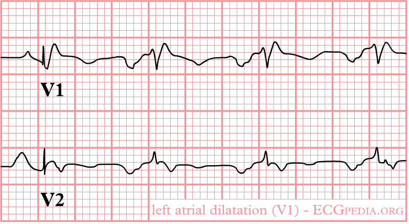
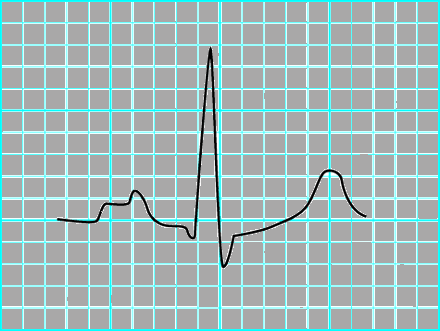
1. LA enlargement: Left atrial enlargement produces a broad, bifid P wave in lead II (P mitrale) and enlarges the terminal negative portion of the P wave in VI.

In lead II following may be seen:

  • Bifid P wave with > 40 ms between the two peaks
  • Total P wave duration > 110 ms


In lead V1 follwing may be seen:

  • Biphasic P wave with terminal negative portion > 40 ms duration
  • Biphasic P wave with terminal negative portion > 1mm deep

Left atrial enlargement as seen in lead V1


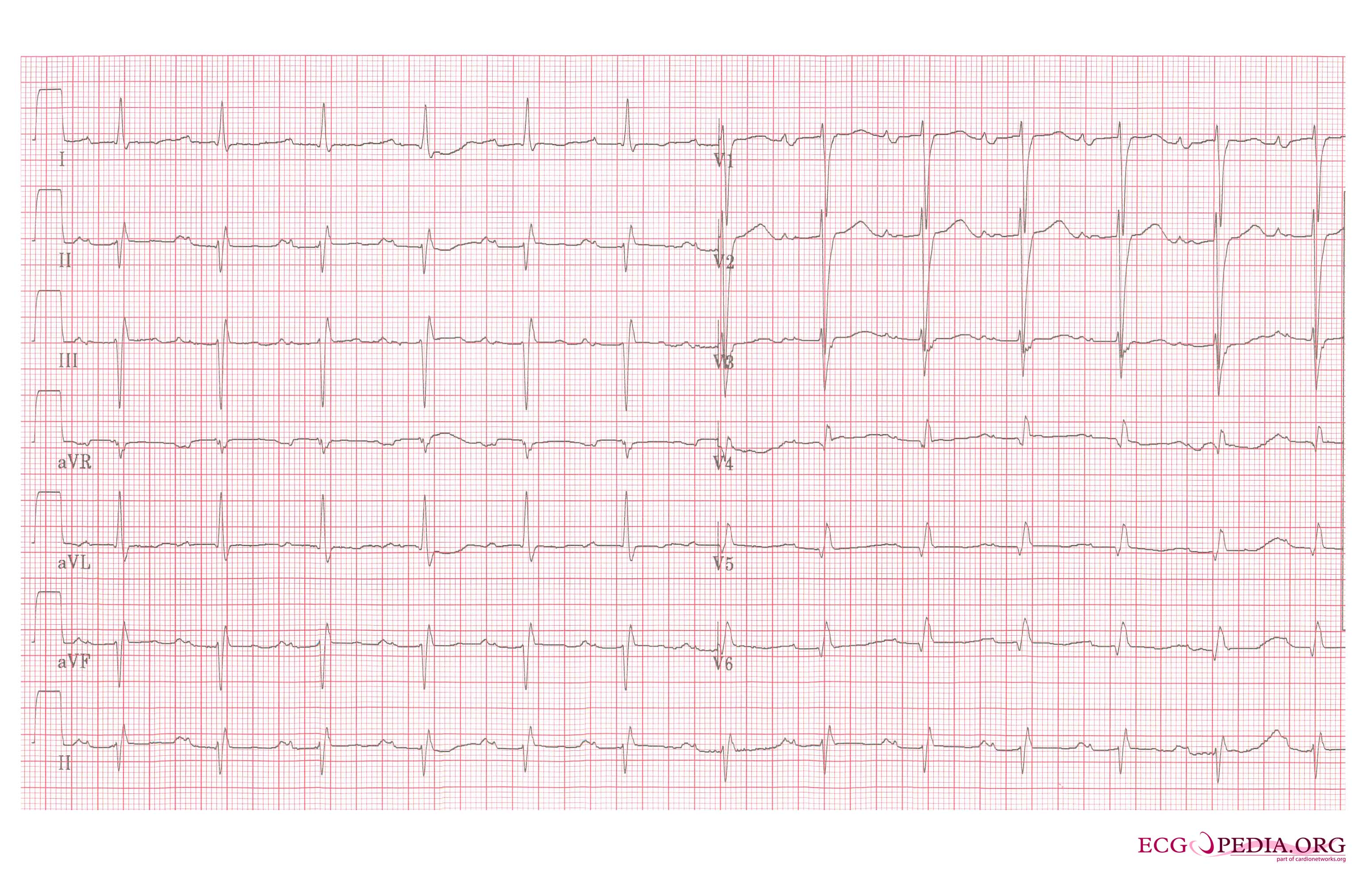
2. Right ventricular hypertrophy: A mean QRS axis in the frontal plane is greater than 80 and an R-to-S ratio of greater than 1 in lead V1.

3. Right axis deviation: mean QRS axis in the frontal plane moves toward the right as pulmonary hypertension worsens.

4. Atrial fibrillation is commonly seen with mitral stenosis: Irregularly irregular rhythm with absence P waves.


Below is an ECG in mitral stenosis
Left atrial enlargement, a 12 lead ECG


References

Template:WikiDoc Sources