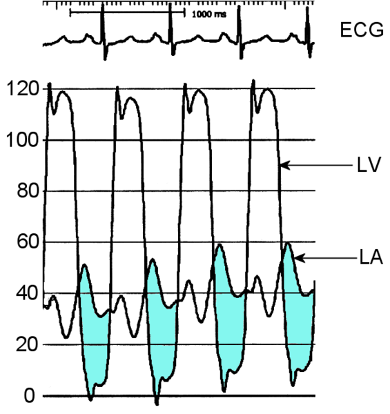
Mitral stenosis cardiac catheterization
Editor-In-Chief: C. Michael Gibson, M.S., M.D. [1]; Associate Editor-In-Chief: Cafer Zorkun, M.D., Ph.D. [2]
Cardiac catheterization
A definitive method of assessing the severity of mitral stenosis is the simultaneous left heart catheterization and right heart catheterization. The right heart catheterization gives the physician the mean pulmonary capillary wedge pressure, which is a reflection of the left atrial pressure. The left heart catheterization, on the other hand, gives the pressure in the left ventricle. By simultaneously taking these pressures, it is possible to determine the gradient between the left atrium and right atrium during ventricular diastole, which is a marker for the severity of mitral stenosis. This method of evaluating mitral stenosis tend to over-estimate the degree of mitral stenosis, however, because of the time lag in the pressure tracings seen on the right heart catheterization and the slow Y descent seen on the wedge tracings. If a trans-septal puncture is made during right heart catheterization, however, the pressure gradient can accurately quantify the severity of mitral stenosis.
