Ischemic stroke MRI: Difference between revisions
Jump to navigation
Jump to search
Aysha Aslam (talk | contribs) No edit summary |
Aysha Aslam (talk | contribs) m (Aysha Aslam moved page Stroke MRI to Ischemic stroke MRI) |
(No difference)
| |
Revision as of 14:44, 7 November 2016
|
Ischemic Stroke Microchapters |
|
Diagnosis |
|---|
|
Treatment |
|
Case Studies |
|
Ischemic stroke MRI On the Web |
|
American Roentgen Ray Society Images of Ischemic stroke MRI |
Editor-In-Chief: C. Michael Gibson, M.S., M.D. [1]Associate Editor(s)-in-Chief: Aysha Anwar, M.B.B.S[2]
Overview
MRI
For diagnosing ischemic stroke in the emergency setting:[1]
- sensitivity= 83%
- specificity= 98%
MRI scan
- sensitivity= 81%
- specificity= 100%
For detecting chronic hemorrhages, an MRI scan is more sensitive.[2]
For the assessment of stable stroke, nuclear medicine scans SPECT and PET/CT may be helpful. SPECT documents cerebral blood flow and PET with FDG isotope the metabolic activity of the neurons.
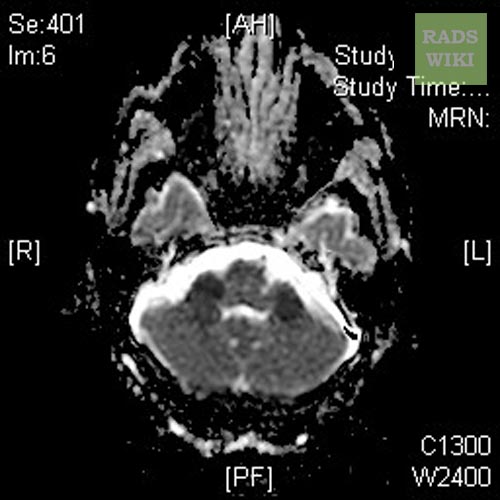
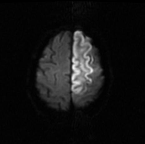
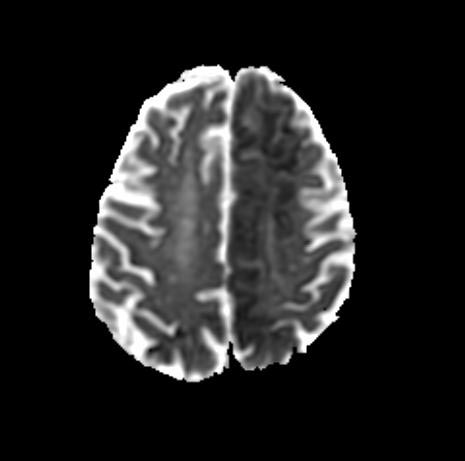
Patient No 1: Change in Mental Status
-
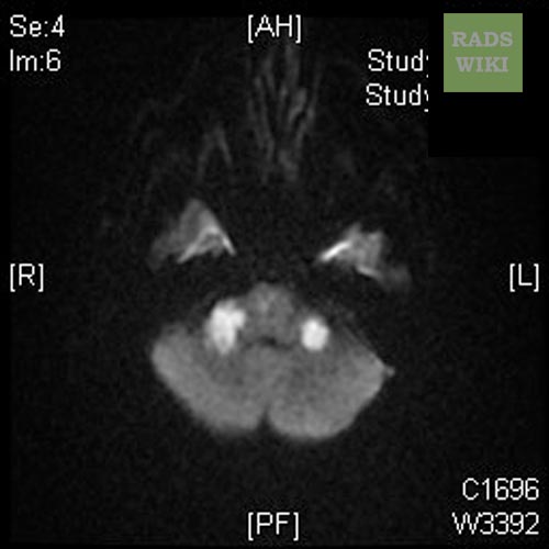
MRI - DWI
-
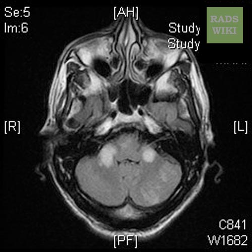
MRI - FLAIR


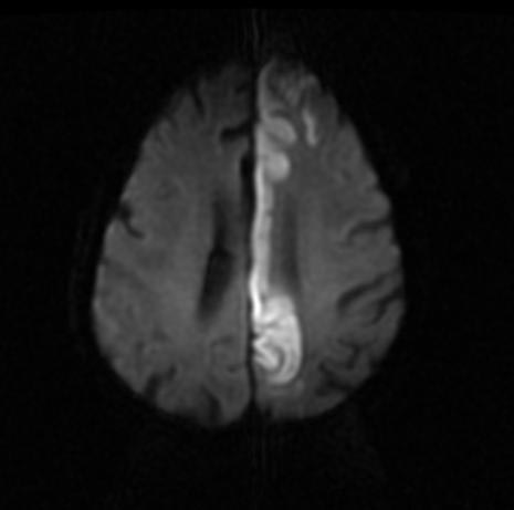
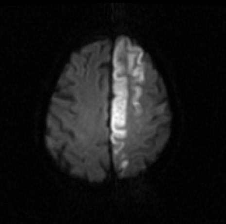
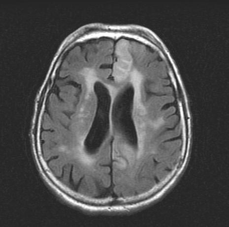
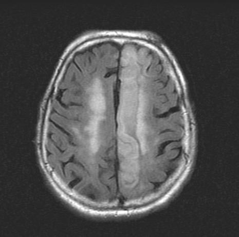
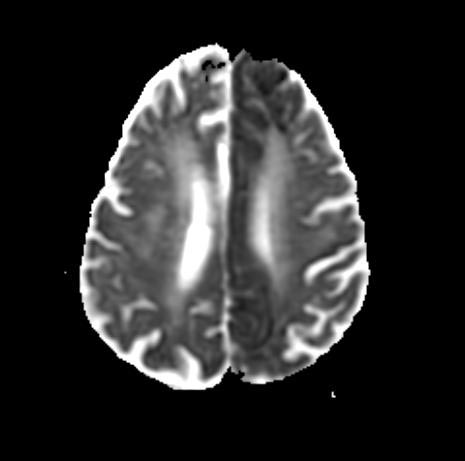
Patient No 2: Left ACA Infarction
-
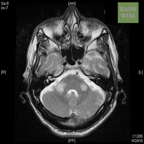
MRI - T2
-
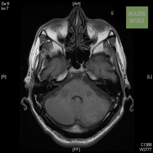
MRI - T1
-
MRI - ADC


-
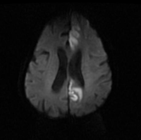
MRI- DWI
-
MRI- DWI


-
MRI- DWI
-
MRI- DWI


-
MRI- FLAIR
-
MRI- FLAIR


-
MRI- ADC
-
MRI- A


References
- ↑ Chalela, J (2007). "Magnetic resonance imaging and computed tomography in emergency assessment of patients with suspected acute stroke: a prospective comparison". Lancet. 369 (9558): 293–8. PMID 17258669. Retrieved 2008-01-22. Unknown parameter
|coauthors=ignored (help) - ↑ Kidwell, C (2004). "Comparison of MRI and CT for detection of acute intracerebral hemorrhage". JAMA. 292 (15): 1823–30. PMID 15494579. Retrieved 2008-01-22. Unknown parameter
|coauthors=ignored (help)