Internal iliac vein
| Cardiology Network |
 Discuss Internal iliac vein further in the WikiDoc Cardiology Network |
| Adult Congenital |
|---|
| Biomarkers |
| Cardiac Rehabilitation |
| Congestive Heart Failure |
| CT Angiography |
| Echocardiography |
| Electrophysiology |
| Cardiology General |
| Genetics |
| Health Economics |
| Hypertension |
| Interventional Cardiology |
| MRI |
| Nuclear Cardiology |
| Peripheral Arterial Disease |
| Prevention |
| Public Policy |
| Pulmonary Embolism |
| Stable Angina |
| Valvular Heart Disease |
| Vascular Medicine |
Editor-In-Chief: C. Michael Gibson, M.S., M.D. [1]
The internal iliac vein (hypogastric vein) begins near the upper part of the greater sciatic foramen, passes upward behind and slightly medial to the hypogastric artery and, at the brim of the pelvis, joins with the external iliac to form the common iliac vein.
Tributaries
With the exception of the fetal umbilical vein which passes upward and backward from the umbilicus to the liver, and the iliolumbar vein which usually joins the common iliac vein, the tributaries of the hypogastric vein correspond with the branches of the hypogastric artery.
| Receives | Description |
| gluteal vein internal pudendal vein obturator veins |
have their origins outside the pelvis; |
| lateral sacral veins | lie in front of the sacrum |
| middle hemorrhoidal vein vesical vein uterine vein vaginal veins |
originate in venous plexuses connected with the pelvic viscera. |
Additional images
-
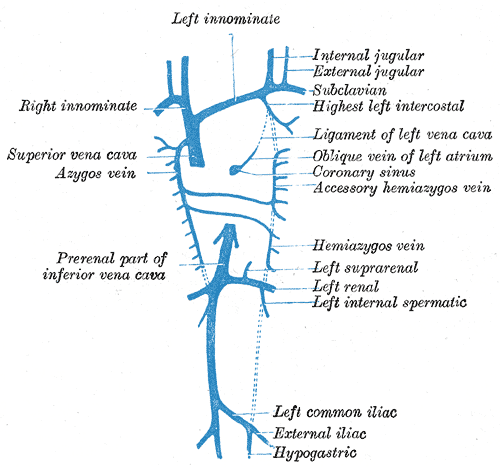
Diagram showing completion of development of the parietal veins.
