Hepatocellular adenoma: Difference between revisions
| Line 74: | Line 74: | ||
<gallery perRow="3"> | <gallery perRow="3"> | ||
Image:Hepatic ademona CT 001.jpg|CT portal venous phase | Image:Hepatic ademona CT 001.jpg|CT portal venous phase: A patient with multiple adenoma | ||
Image:Hepatic ademona CT 002.jpg|CT portal venous phase | Image:Hepatic ademona CT 002.jpg|CT portal venous phase: A patient with multiple adenoma | ||
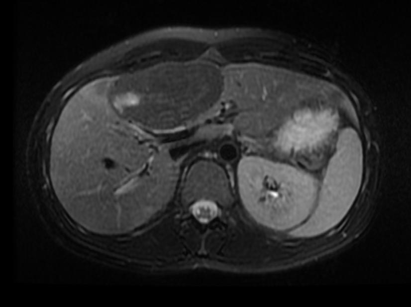
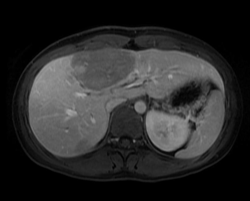
Image:Hepatic ademona MRI 003.jpg|T2 SSFSE | Image:Hepatic ademona MRI 003.jpg|T2 SSFSE: A patient with multiple adenoma | ||
Image:Hepatic ademona MRI 004.jpg|T2 SSFSE | Image:Hepatic ademona MRI 004.jpg|T2 SSFSE: A patient with multiple adenoma | ||
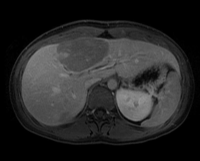
Image:Hepatic ademona MRI 005.jpg|T2 Fat sat | Image:Hepatic ademona MRI 005.jpg|T2 Fat sat: A patient with multiple adenoma | ||
Image:Hepatic ademona MRI 006.jpg|In phase | Image:Hepatic ademona MRI 006.jpg|In phase: A patient with multiple adenoma | ||
Image:Hepatic ademona MRI 007.jpg|Out of phase | Image:Hepatic ademona MRI 007.jpg|Out of phase: A patient with multiple adenoma | ||
Image:Hepatic ademona MRI 008.jpg|T1 fat sat | Image:Hepatic ademona MRI 008.jpg|T1 fat sat: A patient with multiple adenoma | ||
Image:Hepatic ademona MRI 009.jpg|T1 fat sat arterial | Image:Hepatic ademona MRI 009.jpg|T1 fat sat arterial: A patient with multiple adenoma | ||
Image:Hepatic ademona MRI 010.jpg|T1 fat sat arterial | Image:Hepatic ademona MRI 010.jpg|T1 fat sat arterial: A patient with multiple adenoma | ||
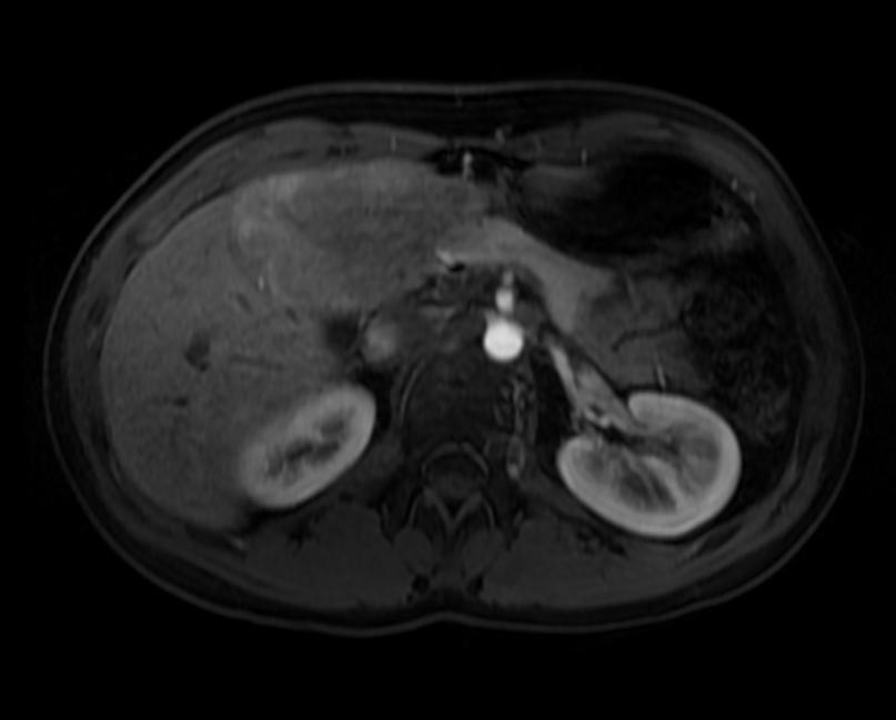
Image:Hepatic ademona MRI 011.jpg|T1 fat sat delayed | Image:Hepatic ademona MRI 011.jpg|T1 fat sat delayed: A patient with multiple adenoma | ||
Image:Hepatic ademona MRI 012.jpg|T1 fat sat delayed | Image:Hepatic ademona MRI 012.jpg|T1 fat sat delayed: A patient with multiple adenoma | ||
</gallery> | </gallery> | ||
Revision as of 02:12, 7 April 2009
| Hepatocellular adenoma | |
| ICD-O: | Template:ICDO |
|---|---|
| DiseasesDB | 5726 |
| eMedicine | med/48 |
| MeSH | D018248 |
Editor-In-Chief: C. Michael Gibson, M.S., M.D. [1]
Contributors: Cafer Zorkun M.D., PhD.
Please Take Over This Page and Apply to be Editor-In-Chief for this topic: There can be one or more than one Editor-In-Chief. You may also apply to be an Associate Editor-In-Chief of one of the subtopics below. Please mail us [2] to indicate your interest in serving either as an Editor-In-Chief of the entire topic or as an Associate Editor-In-Chief for a subtopic. Please be sure to attach your CV and or biographical sketch.
Overview
Hepatocellular adenoma, also hepatic adenoma, or rarely hepadenoma, is an uncommon benign liver tumour which is associated with the use of types of hormonal contraception with a high estrogen content.[1]
Diagnosis
MRI is the most useful investigation in the diagnosis and workup.[2]
Diagnostic Findings
Ultrasonography
- Color Doppler US may demonstrate peripheral peritumoral vessels and intratumoral vessels that typically have a flat continuous or, less commonly, triphasic waveform. *These Doppler US features are reported to be absent in the vessels within focal nodular hyperplasia and may be useful in distinguishing the two disease entities. *Nevertheless, most adenomas are not specifically diagnosed at US and are usually further evaluated with CT or other imaging modalities.
Computed Tomography
- Fat or hemorrhage can easily be identified on unenhanced images, and delayed-phase images demonstrate the tendency for fibrotic components to enhance and retain contrast material.
- Because adenomas consist almost entirely of uniform hepatocytes and a variable number of Kupffer cells, most adenomas are nearly isoattenuating relative to normal liver on unenhanced, portal venous–phase, and delayed-phase images.
- In patients with fatty liver, adenomas are hyperattenuating at all phases of contrast enhancement and on unenhanced images as well.
- Small hepatocellular adenomas enhance rapidly and are hyperattenuating relative to the liver.
- Excluding lesions with acute or old tumor hemorrhage and fat deposition, hepatocellular adenoma demonstrated homogeneous or nearly homogeneous enhancement in approx 80% of cases.
- The enhancement usually does not persist in adenomas because of arteriovenous shunting.
- Larger hepatocellular adenomas may be more heterogeneous than smaller lesions, and their CT appearance is less specific.
Magnetic Resonance Imaging
- On T1-weighted MR images, hepatocellular adenomas have been variously described as hyperintense, isointense, and hypointense lesions.
- It has been reported that 47%–74% of hepatocellular adenomas are predominantly hyperintense relative to liver on T2-weighted images; this is due to prolonged T2 and is consistent with findings in other hepatic tumors.
- Some lesions are hypointense and isointense on T2-weighted images.
- Most lesions are heterogeneous, demonstrating a combination of hyper- and hypointensity on T2-weighted images relative to hemorrhage and necrosis.
- Dynamic gadolinium-enhanced gradient-echo MR imaging, like dynamic CT, can be used to demonstrate early arterial enhancement that reflects the presence of subcapsular feeding vessels.
- Adenomas usually do not show uptake of superparamagnetic iron oxide particles, resulting in decreased signal intensity on T2-weighted images.
- After injection of a hepatocellular-specific contrast agent such as gadolinium benzyloxypropionictetraacetate (Gd-BOPTA) there is usually no substantial uptake.
Nuclear Scintigraphy
- Compared with normal liver, adenomas usually show absent or decreased uptake of Tc-99m sulfur colloid, reflecting the decreased number or function of Kupffer cells.
-
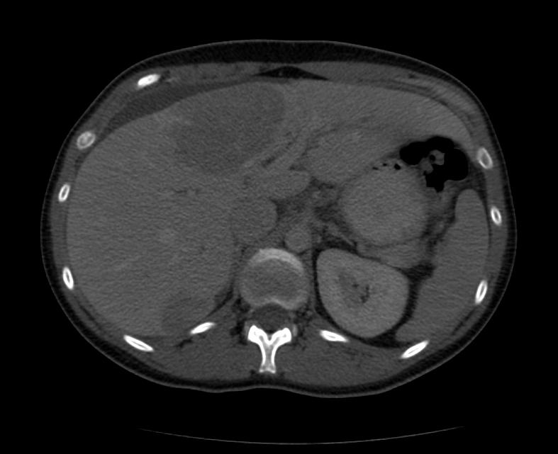
CT portal venous phase: A patient with multiple adenoma
-
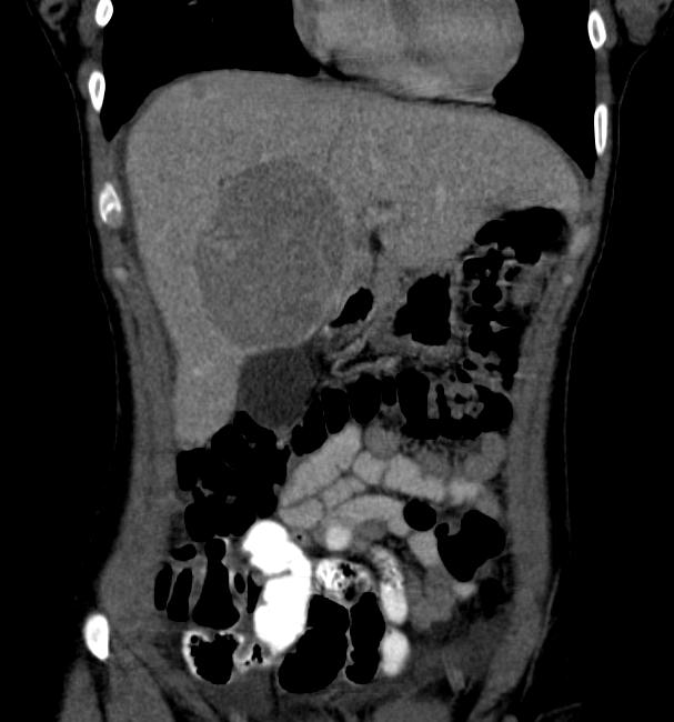
CT portal venous phase: A patient with multiple adenoma
-
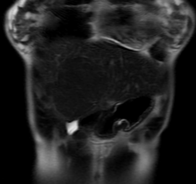
T2 SSFSE: A patient with multiple adenoma
-
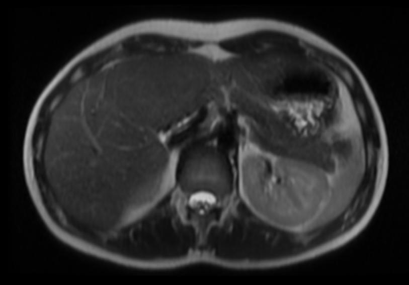
T2 SSFSE: A patient with multiple adenoma
-
T2 Fat sat: A patient with multiple adenoma
-
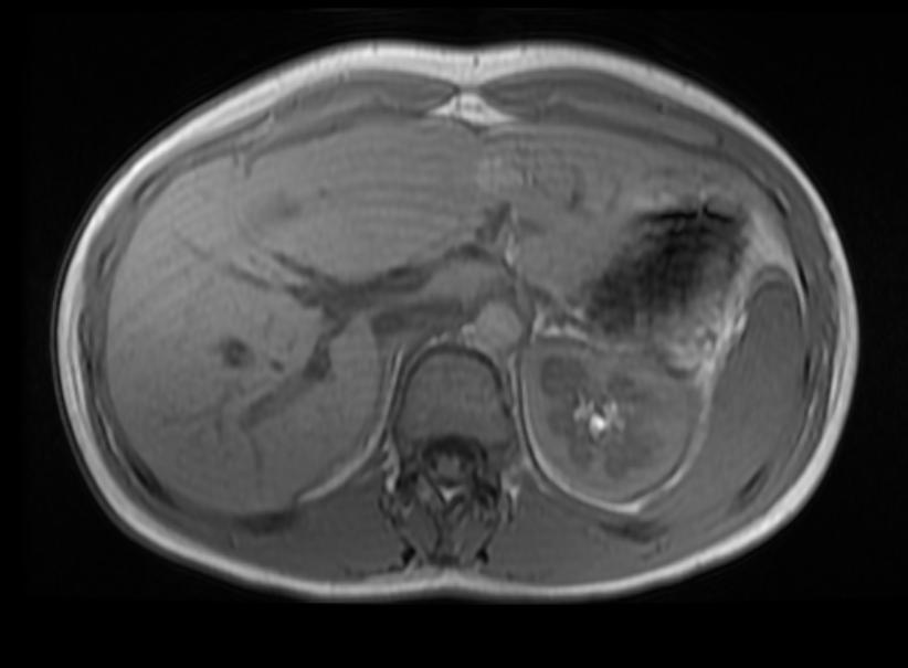
In phase: A patient with multiple adenoma
-
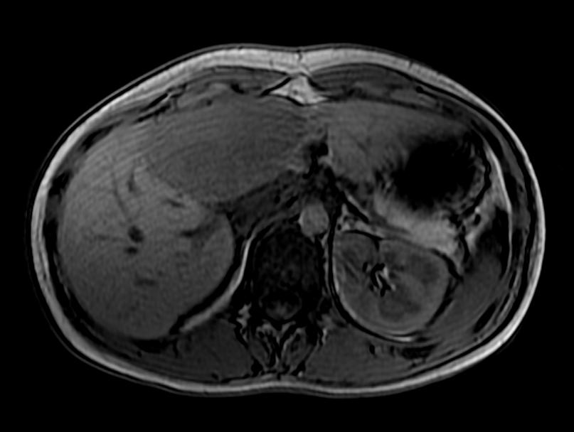
Out of phase: A patient with multiple adenoma
-
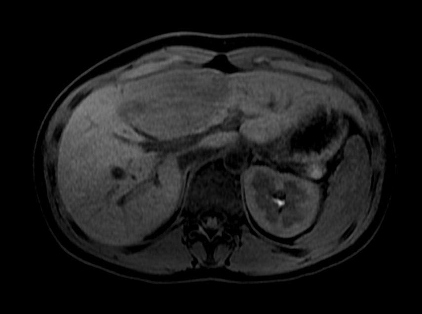
T1 fat sat: A patient with multiple adenoma
-
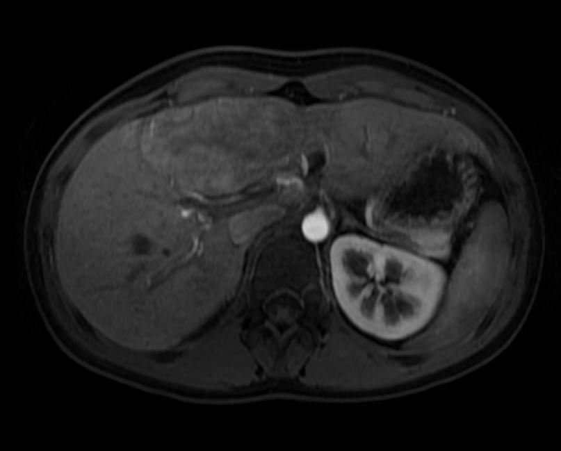
T1 fat sat arterial: A patient with multiple adenoma
-
T1 fat sat arterial: A patient with multiple adenoma
-
T1 fat sat delayed: A patient with multiple adenoma
-
T1 fat sat delayed: A patient with multiple adenoma


Treatment
All hepatocellular adenoma should be surgically resected, because of the risk of rupture causing bleeding and because they may contain malignant foci.[3]
References
- ↑ Rooks J, Ory H, Ishak K, Strauss L, Greenspan J, Hill A, Tyler C (1979). "Epidemiology of hepatocellular adenoma. The role of oral contraceptive use". JAMA. 242 (7): 644–8. PMID 221698.
- ↑ Hussain S, van den Bos I, Dwarkasing R, Kuiper J, den Hollander J (2006). "Hepatocellular adenoma: findings at state-of-the-art magnetic resonance imaging, ultrasound, computed tomography and pathologic analysis". Eur Radiol. 16 (9): 1873–86. PMID 16708218.
- ↑ Toso C, Majno P, Andres A, Rubbia-Brandt L, Berney T, Buhler L, Morel P, Mentha G (2005). "Management of hepatocellular adenoma: solitary-uncomplicated, multiple and ruptured tumors". World J Gastroenterol. 11 (36): 5691–5. PMID 16237767.Full text