Hemopericardium: Difference between revisions
| Line 30: | Line 30: | ||
[http://www.peir.net Images courtesy of Professor Peter Anderson DVM PhD and published with permission © PEIR, University of Alabama at Birmingham, Department of Pathology] | [http://www.peir.net Images courtesy of Professor Peter Anderson DVM PhD and published with permission © PEIR, University of Alabama at Birmingham, Department of Pathology] | ||
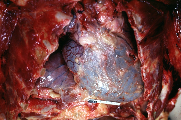
Image:Hemopericardium 001.jpg|Hemopericardium: Gross, an excellent in situ view | [[Image:Hemopericardium 001.jpg|Hemopericardium: Gross, an excellent in situ view]] | ||
<br clear="left"/> | <br clear="left"/> | ||
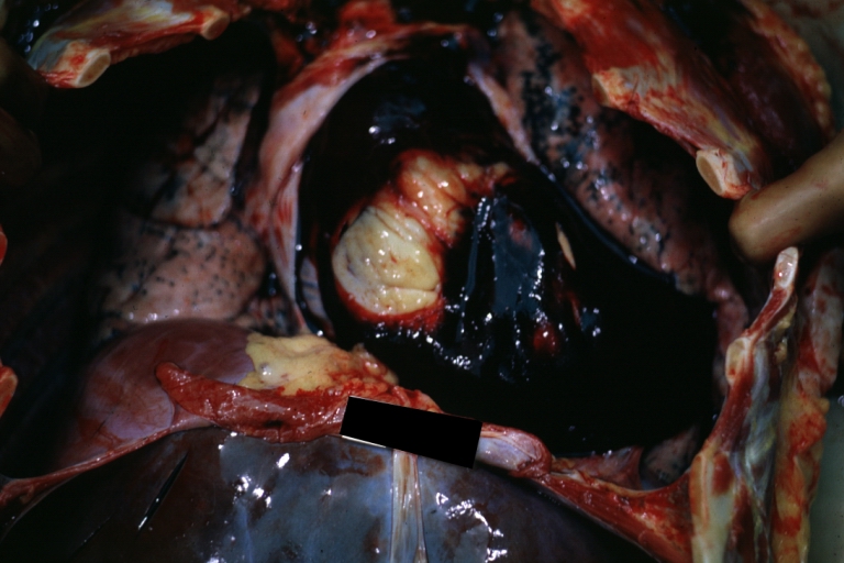
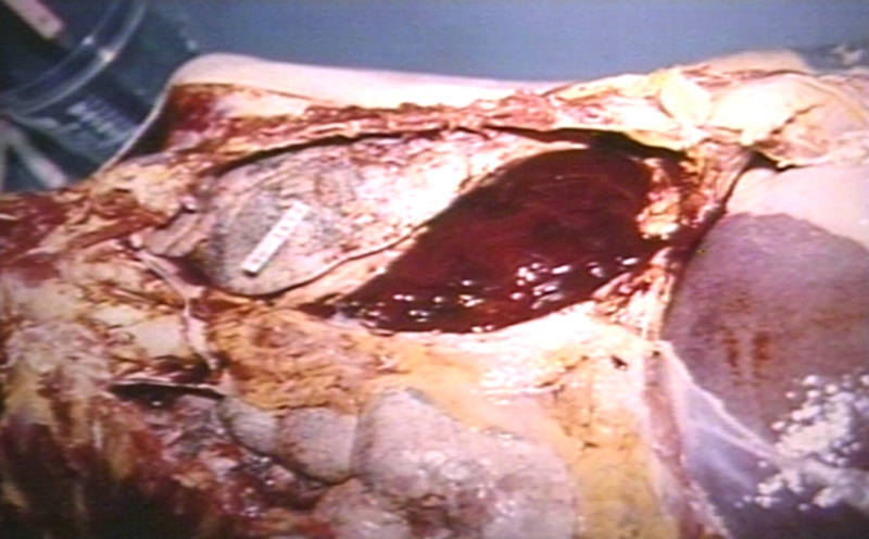
Image:Hemopericardium 002.jpg|Hemopericardium: Gross, in situ, unopened pericardium (a very good example) | [[Image:Hemopericardium 002.jpg|Hemopericardium: Gross, in situ, unopened pericardium (a very good example)]] | ||
<br clear="left"/> | <br clear="left"/> | ||
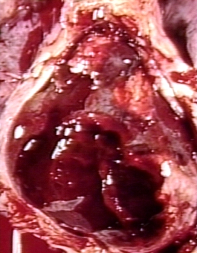
Image:Hemopericardium 003.jpg|Hemopericardium: Gross, natural color, heart in situ with opened pericardium and filled with red blood clot (quite good example) dissecting aneurysm | [[Image:Hemopericardium 003.jpg|Hemopericardium: Gross, natural color, heart in situ with opened pericardium and filled with red blood clot (quite good example) dissecting aneurysm]] | ||
<br clear="left"/> | <br clear="left"/> | ||
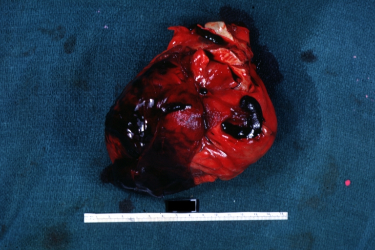
Image:Hemopericardium 004.jpg|Hemopericardium due to Needle Puncture: Gross, natural color, external view of heart covered by blood | [[Image:Hemopericardium 004.jpg|Hemopericardium due to Needle Puncture: Gross, natural color, external view of heart covered by blood ]] | ||
<br clear="left"/> | <br clear="left"/> | ||
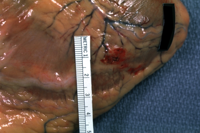
Image:Hemopericardium 005.jpg|Needle Puncture Mark in Epicardium: Gross, natural color, close-up of needle puncture marks tap resulted in hemopericardium | [[Image:Hemopericardium 005.jpg|Needle Puncture Mark in Epicardium: Gross, natural color, close-up of needle puncture marks tap resulted in hemopericardium ]] | ||
<br clear="left"/> | <br clear="left"/> | ||
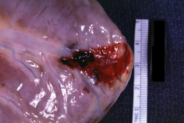
Image:Hemopericardium 006.jpg|Hemopericardium: Hemopericardium caused by pericardiocentesis: Gross, natural color, close-up view of apex of the heart. Needle apparently entered the distal posterior descending artery. | [[Image:Hemopericardium 006.jpg|Hemopericardium: Hemopericardium caused by pericardiocentesis: Gross, natural color, close-up view of apex of the heart. Needle apparently entered the distal posterior descending artery.]] | ||
<br clear="left"/> | <br clear="left"/> | ||
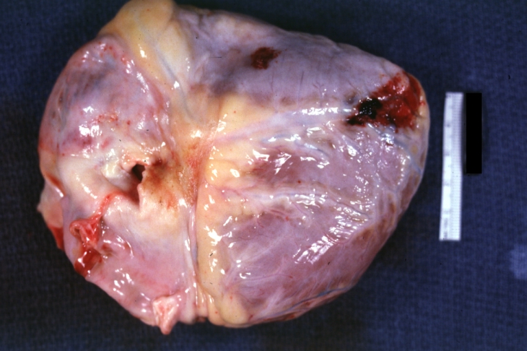
Image:Hemopericardium 007.jpg|Hemopericardium: Hemopericardium caused by pericardiocentesis: Gross, natural color, view of apex of the heart. Needle apparently entered the distal posterior descending artery | [[Image:Hemopericardium 007.jpg|Hemopericardium: Hemopericardium caused by pericardiocentesis: Gross, natural color, view of apex of the heart. Needle apparently entered the distal posterior descending artery]] | ||
<br clear="left"/> | <br clear="left"/> | ||
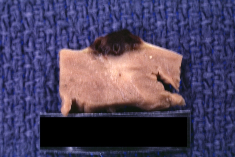
Image:Hemopericardium 008.jpg|Hemopericardium: Hemopericardium due to pericardiocentesis: Gross, fixed tissue, close-up view of slice through distal posterior descending artery showing periarterial hemorrhage | [[Image:Hemopericardium 008.jpg|Hemopericardium: Hemopericardium due to pericardiocentesis: Gross, fixed tissue, close-up view of slice through distal posterior descending artery showing periarterial hemorrhage]] | ||
<br clear="left"/> | <br clear="left"/> | ||
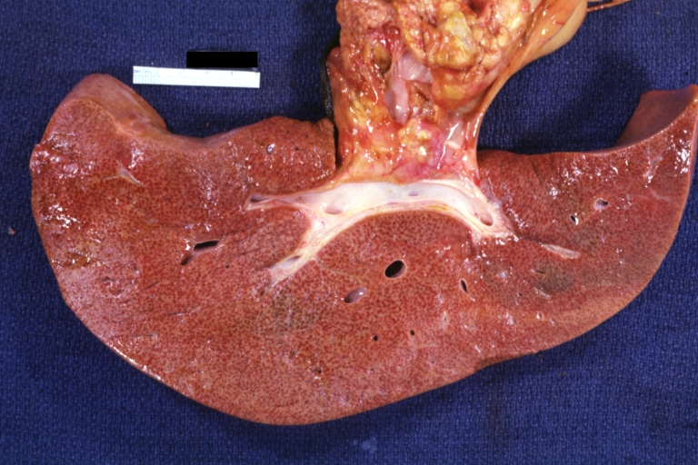
Image:Hemopericardium 009.jpg|Hemopericardium: Liver: Gross, natural color, typical shock liver case of death due to hemopericardium secondary to pericardiocentesis | [[Image:Hemopericardium 009.jpg|Hemopericardium: Liver: Gross, natural color, typical shock liver case of death due to hemopericardium secondary to pericardiocentesis]] | ||
<br clear="left"/> | <br clear="left"/> | ||
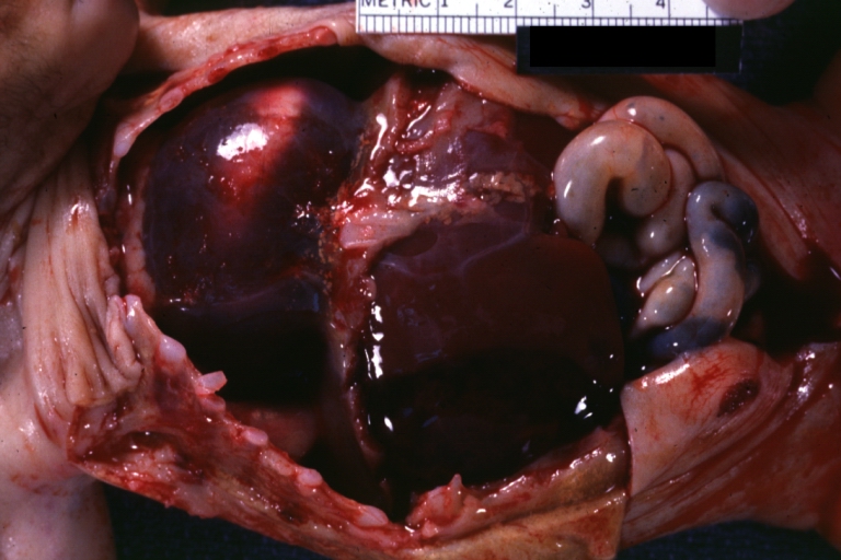
Image:Hemopericardium 010.jpg|Hemopericardium in newborn: Gross, natural color, opened body with large collection blood in pericardial sac. Cause uncertain. A 26 week premature with hyaline membrane disease and DIC | [[Image:Hemopericardium 010.jpg|Hemopericardium in newborn: Gross, natural color, opened body with large collection blood in pericardial sac. Cause uncertain. A 26 week premature with hyaline membrane disease and DIC]] | ||
<br clear="left"/> | <br clear="left"/> | ||
Image:Hemopericardium 011.jpg|Hemopericardium: Myocardial Infarction and Ventricular Rupture | [[Image:Hemopericardium 011.jpg|Hemopericardium: Myocardial Infarction and Ventricular Rupture]] | ||
<br clear="left"/> | <br clear="left"/> | ||
Image:Hemopericardium 012.jpg|Hemopericardium: Infarct rupture after 7 days of chest pain onset. | [[Image:Hemopericardium 012.jpg|Hemopericardium: Infarct rupture after 7 days of chest pain onset.]] | ||
<br clear="left"/> | <br clear="left"/> | ||
Image:Hemopericardium 013.jpg|Hemopericardium in dissecting aneurysm: Gross, heart with root of aorta to show hemorrhage into pericardium (very good example) | [[Image:Hemopericardium 013.jpg|Hemopericardium in dissecting aneurysm: Gross, heart with root of aorta to show hemorrhage into pericardium (very good example)]] | ||
<br clear="left"/> | <br clear="left"/> | ||
Revision as of 21:16, 6 March 2009
| Hemopericardium | |
 | |
|---|---|
| Hemopericardium: Gross, an excellent in situ view. Image courtesy of Professor Peter Anderson DVM PhD and published with permission © PEIR, University of Alabama at Birmingham, Department of Pathology | |
| ICD-10 | I23.0, I31.2, S26.0 |
| ICD-9 | 423.0, 860.2 |
|
WikiDoc Resources for Hemopericardium |
|
Articles |
|---|
|
Most recent articles on Hemopericardium Most cited articles on Hemopericardium |
|
Media |
|
Powerpoint slides on Hemopericardium |
|
Evidence Based Medicine |
|
Clinical Trials |
|
Ongoing Trials on Hemopericardium at Clinical Trials.gov Trial results on Hemopericardium Clinical Trials on Hemopericardium at Google
|
|
Guidelines / Policies / Govt |
|
US National Guidelines Clearinghouse on Hemopericardium NICE Guidance on Hemopericardium
|
|
Books |
|
News |
|
Commentary |
|
Definitions |
|
Patient Resources / Community |
|
Patient resources on Hemopericardium Discussion groups on Hemopericardium Patient Handouts on Hemopericardium Directions to Hospitals Treating Hemopericardium Risk calculators and risk factors for Hemopericardium
|
|
Healthcare Provider Resources |
|
Causes & Risk Factors for Hemopericardium |
|
Continuing Medical Education (CME) |
|
International |
|
|
|
Business |
|
Experimental / Informatics |
Editor-In-Chief: C. Michael Gibson, M.S., M.D. [1]
Associate Editor-In-Chief: Cafer Zorkun, M.D., Ph.D. [2]
Please Take Over This Page and Apply to be Editor-In-Chief for this topic: There can be one or more than one Editor-In-Chief. You may also apply to be an Associate Editor-In-Chief of one of the subtopics below. Please mail us [3] to indicate your interest in serving either as an Editor-In-Chief of the entire topic or as an Associate Editor-In-Chief for a subtopic. Please be sure to attach your CV and or biographical sketch.
Overview
Hemopericardium refers to blood in the pericardial sac of the heart. It is a cause of pericardial effusion, and can also cause cardiac tamponade.[1]
The condition can be caused by trauma,[2] but it has also been observed in patients on anticoagulant therapy.[3][4]
Pathological Findings
References
- ↑ "Forensic Pathology".
- ↑ Krejci CS, Blackmore CC, Nathens A (2000). "Hemopericardium: an emergent finding in a case of blunt cardiac injury". AJR Am J Roentgenol. 175 (1): 250. PMID 10882282. Unknown parameter
|month=ignored (help) - ↑ Katis PG (2005). "Atraumatic hemopericardium in a patient receiving warfarin therapy for a pulmonary embolus". CJEM. 7 (3): 168–70. PMID 17355673. Unknown parameter
|month=ignored (help) - ↑ Hong YC, Chen YG, Hsiao CT, Kuan JT, Chiu TF, Chen JC (2007). "Cardiac tamponade secondary to haemopericardium in a patient on warfarin". Emerg Med J. 24 (9): 679–80. doi:10.1136/emj.2007.049643. PMID 17711963. Unknown parameter
|month=ignored (help)
Template:Heart diseases Template:Injuries, other than fractures, dislocations, sprains and strains Template:Hemodynamics Template:SIB
