Giant cell tumor of bone: Difference between revisions
| Line 49: | Line 49: | ||
:*Local recurrence rate of giant cell tumor of bone is 10 to 40%. | :*Local recurrence rate of giant cell tumor of bone is 10 to 40%. | ||
:*Recurrence rates are higher when the tumor bone arises at a surgical inaccessible location locations such as [[spine]] and [[sacrum]]. | :*Recurrence rates are higher when the tumor bone arises at a surgical inaccessible location locations such as [[spine]] and [[sacrum]]. | ||
*Metastasis | |||
:*Giant cell tumor of bone may occasionally metastasize to vital organs such as the lung.<ref name="MuheremuNiu2014">{{cite journal|last1=Muheremu|first1=Aikeremujiang|last2=Niu|first2=Xiaohui|title=Pulmonary metastasis of giant cell tumor of bones|journal=World Journal of Surgical Oncology|volume=12|issue=1|year=2014|pages=261|issn=1477-7819|doi=10.1186/1477-7819-12-261}}</ref> | |||
==Diagnosis== | ==Diagnosis== | ||
Revision as of 16:32, 11 March 2016
For patient information, click here Editor-In-Chief: C. Michael Gibson, M.S., M.D. [1]
Synonyms and keywords: Osteoclastoma; giant cell myeloma; giant cell tumor
Overview
Historical Perspective
Pathophysiology
Giant cell tumor of bone occurs commonly in the distal femur, the proximal tibia, the distal radius and the sacrum. Giant cell tumors (GCT) usually prefers the epiphyses of long bones.[1]
Microscopic Pathology
- Giant cell tumor of bone is characterized by the presence of numerous Cathepsin-K producing, CD33 +, CD14 - multinucleated osteoclast-like giant cells and plump spindle-shaped stromal cells that represent the main proliferating cell population.
- The spindle-shaped mononuclear cells are believed to represent the neoplastic population and are characterized at the cytogenetic level by telomeric associations and a peculiar telomere-protecting capping mechanism.
- Areas of regressive change such as necrosis or fibrosis as well as extensive hemorrhage are frequently present.
- It recurs from time to time and rates between 25–50% have been reported. In very rare cases, a malignant change may occur.
Causes
Epidemiology and Demographics
Prevalence
- Giant cell tumor of bone accounts for 4-5% of primary bone tumors and 18.2% of benign bone tumors [2].
Age
- Giant cell tumor of bone almost invariably (97-99%) occur when the growth plate has closed and are therefore typically observed in early adulthood, with 80% of cases reported between the ages of 20 and 50, with a peak incidence between 20 and 30.[3]
Gender
- Giant cell tumor of bone is more common in females with the rate of growth enhanced in pregnancy. Female to male ratio 1.5:1.[4]
Risk Factors
Differentiating Giant cell tumor of bone from other Diseases
A number of tumors have giant cells, but are not true benign giant cell tumors. These include
- Aneurysmal bone cyst
- Chondroblastoma
- Simple bone cyst
- Osteoid osteoma
- Osteoblastoma
- Osteosarcoma
- Giant cell reparative granuloma
- Brown tumor of hyperparathyroidism.
Natural History, Complications and Prognosis
Complications
- Malignant transformation
- Malignant transformation is far more common in men (M:F of ~3:1)
- Recurrence
- Metastasis
- Giant cell tumor of bone may occasionally metastasize to vital organs such as the lung.[4]
Diagnosis
Staging
History and Symptoms
- Patients usually present with pain and limited range of motion caused by tumor's proximity to the joint space.
- There may be swelling as well, if the tumor has been growing for a long time.
- Some patients may be asymptomatic until they develop a pathologic fracture at the site of the tumor.
Physical Examination
Laboratory Findings
X Ray
General radiographic features include:
- Narrow zone of transition: a broader zone of transition is seen in more aggressive giant cell tumors.
- No surrounding sclerosis: 80-85%
- Overlying cortex is thinned, expanded or deficient
- Periosteal reaction is only observed in 10-30% of cases
- Soft tissue mass is not infrequent
- Pathological fracture may be present
- No matrix calcification/mineralisation
On x-ray, giant cell tumors (GCTs) have a metaepiphyseal location and grow to the articular surface of the involved bone [5]. They are distinguishable from other bony tumors in that GCTs usually have a non-sclerotic and sharply defined border. Because giant cell tumors are known to metastasize, when the diagnosis of giant cell tumor is suspected, a chest x-ray or CT may be needed.
(Images courtesy of RadsWiki)
-
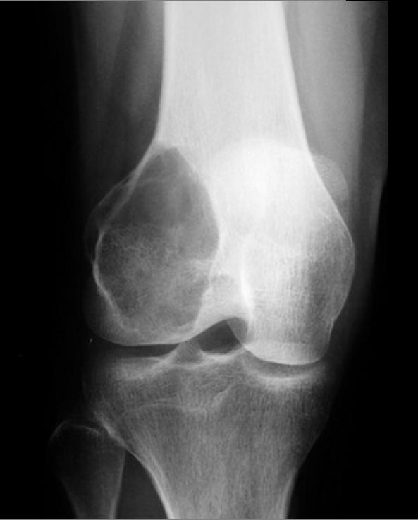
Giant cell tumor: Distal part of the femur
-
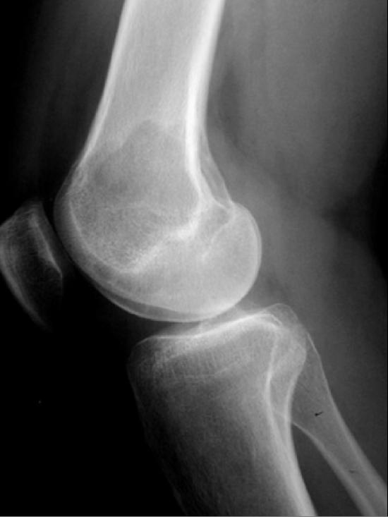
Giant cell tumor: Distal part of the femur


CT
MRI
Typical signal characteristics on MRI of giant cell tumor of bone include:
T1:
- Low to intermediate solid component
- Low signal periphery
- Solid components enhance, helping distinguish GCT with ABC from pure ABC 3-4
- Some enhancement may also be seen in adjacent bone marrow
T2:
- Heterogenous high signal with areas of low signal intensity (variable) due to haemosiderin or fibrosis
- If an ABC component present, then fluid-fluid levels can be observed
- High signal in adjacent bone marrow thought to represent inflammatory edema
T1 C+ (Gd):
- Solid components will enhance, helping differentiate from ABCs
Treatment
Medical Therapy
Surgery
References
- ↑ Shrivastava, Sandeep; Nawghare, Shishir P; Kolwadkar, Yogesh; Singh, Pradeep (2008). "Giant cell tumour in the diaphysis of radius – a report". Cases Journal. 1 (1): 106. doi:10.1186/1757-1626-1-106. ISSN 1757-1626.
- ↑ Gamberi G, Serra M, Ragazzini P, Magagnoli G, Pazzaglia L, Ponticelli F, Ferrari C, Zanasi M, Bertoni F, Picci P, Benassi MS (2003). "Identification of markers of possible prognostic value in 57 giant cell tumors of bone". Oncology Reports. 10 (2): 351–6. PMID 12579271. Retrieved 2012-01-18.
- ↑ Giant cell tumor of bone.Dr Henry Knipe and Dr Behrang Amini et al.Radiopaedia.org 2015.http://radiopaedia.org/articles/giant-cell-tumour-of-bone.Accessed on March 11, 2016
- ↑ 4.0 4.1 Muheremu, Aikeremujiang; Niu, Xiaohui (2014). "Pulmonary metastasis of giant cell tumor of bones". World Journal of Surgical Oncology. 12 (1): 261. doi:10.1186/1477-7819-12-261. ISSN 1477-7819.
- ↑ Murphey MD, Nomikos GC, Flemming DJ, Gannon FH, Temple HT, Kransdorf MJ (2001). "From the archives of AFIP. Imaging of giant cell tumor and giant cell reparative granuloma of bone: radiologic-pathologic correlation". Radiographics : a Review Publication of the Radiological Society of North America, Inc. 21 (5): 1283–309. PMID 11553835. Retrieved 2012-01-18.