Eosinophilic esophagitis overview

Jump to navigation Jump to search

Eosinophilic Esophagitis Microchapters

Home

Patient Information

Overview

Historical Perspective

Classification

Pathophysiology

Causes

Differentiating Eosinophilic Esophagitis from other Diseases

Epidemiology and Demographics

Risk Factors

Screening

Natural History, Complications and Prognosis

Diagnosis

History and Symptoms

Physical Examination

Laboratory Findings

X Ray

CT

MRI

Other Imaging Findings

Other Diagnostic Studies

Treatment

Medical Therapy

Surgery

Primary Prevention

Secondary Prevention

Future or Investigational Therapies

Case Studies

Case #1

Eosinophilic esophagitis overview On the Web

Most recent articles

Most cited articles

Review articles

CME Programs

Powerpoint slides

Images

American Roentgen Ray Society Images of Eosinophilic esophagitis overview

All Images
X-rays
Echo & Ultrasound
CT Images
MRI

Ongoing Trials at Clinical Trials.gov

US National Guidelines Clearinghouse

NICE Guidance

FDA on Eosinophilic esophagitis overview

CDC on Eosinophilic esophagitis overview

Eosinophilic esophagitis overview in the news

Blogs on Eosinophilic esophagitis overview

Directions to Hospitals Treating Eosinophilic esophagitis

Risk calculators and risk factors for Eosinophilic esophagitis overview

Editor-In-Chief: C. Michael Gibson, M.S., M.D. [1]

Overview

Eosinophilic esophagitis is an allergic inflammatory condition of the esophagus. Symptoms are chest pain or heartburn and occasionally dysphagia, or difficulty in swallowing. The disease was first described in children but occurs in adults as well.
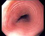
Endoscopic image of esophagus in a case of eosinophilic esophagitis. Concentric rings are termed trachealization of the esophagus.

Diagnosis is obtained during an upper GI endoscopy where biopsies are taken of the esophagus. At the time of endoscopy, ridges or furrows may be seen in the esophagus wall. Sometimes, multiple rings may occur in the esophagus, leading to the term "multi-ring esophagus" or "feline esophagus" due to the similarity in the rings of the cat esophagus. A high number of eosinophils are seen on microscopic examination of the biopsy specimens. Skin testing can help identify which foods might contribute to this disease, but often skin testing implicates foods that are not involved. Common allergens in the GI tract are cow's milk, soy, egg and wheat.

Barium swallow of the esophagus, showing multiple rings associated with eosinophilic esophagitis.

Treatment strategies include removal of the offending food, proton pump inhibitors to decrease acidity in the stomach that may reflux into the esophagus, inhaled steroid puffers taken orally and swallowed, anti-histamines, H2-receptor blockers such as cimetidine, leukotriene modifiers such as montelukast, and, in investigational reports, the anti-IL5 monoclonal antibody mepolizumab. Refractory patients may require oral steroid medications.

H&E stain of esophagus biopsy showing eosinophilic esophagitis, manifested by an infiltration of eosinophils in the lamina propria

Historical Perspective

Classification

Pathophysiology

Causes

Differentiating Eosinophilic esophagitis overview from Other Diseases

Epidemiology and Demographics

Risk Factors

Screening

Natural History, Complications, and Prognosis

Natural History

Complications

Prognosis

Diagnosis

Diagnostic Criteria

History and Symptoms

Physical Examination

Laboratory Findings

Imaging Findings

Other Diagnostic Studies

Treatment

Medical Therapy

Surgery

Prevention

References

Template:Gastroenterology

Template:WH Template:WS