Conjunctivitis pathophysiology
Editor-In-Chief: C. Michael Gibson, M.S., M.D. [5] Associate Editor(s)-in-Chief: Sara Mehrsefat, M.D. [6]
|
Conjunctivitis Microchapters |
|
Diagnosis |
|---|
|
Treatment |
|
Case Studies |
|
Conjunctivitis pathophysiology On the Web |
|
American Roentgen Ray Society Images of Conjunctivitis pathophysiology |
|
Risk calculators and risk factors for Conjunctivitis pathophysiology |
Overview
Conjunctivitis is defined as inflammation of bulbar and/or palpebral conjunctiva. Conjunctivitis has many etiologies, but majority of cases can be caused by allergies, viruses, or bacteria. Viral conjunctivitis, typically caused by Adenovirus, is a common, self-limiting condition. Bacterial conjunctivitis has many etiologies, the most common include: Staphylococcus, Streptococcus, Corynebacterium, Haemophilus, Pseudomonas, and Moraxella species.[1] Allergic conjunctivitis may occur seasonally when pollen counts are high, and this type of conjunctivitis is a common occurrence in people who have other signs of allergic disease.[2]
Pathophysiology
Infective conjunctivitis is an infection of the conjunctiva either cause by viruses, or bacteria . Both the palpebral and the bulbar ocular conjunctival surfaces are usually affected and typically become red, and inflamed. Bacterial, and viral conjunctivitis is spread from:[1][2]
- Direct contact with the infected person’s eye drainage or drainage from the person’s cough, sneeze, or runny nose
- Contact with the infected person’s fingers, hands or objects (eye makeup applicators, towels, shared eye medications) that may have the infected person’s drainage on them
- Adjacent infectious sites (rubbing of the eyes)
Any change in the host defense, or in the species of normal flora of the eye (such as Streptococci, Staphylococci, and Corynebacteria) can lead to clinical infection and conjunctivitis. Change in the normal flora can occurred by:[3]
- External contamination
- Contact lens wear
- Swimming
Viral conjunctivitis that is typically caused by adenovirus is a common, self-limiting condition. Viral conjunctivitis is highly contagious, and patients should avoid direct or indirect contact with other healthy individuals. Acute hemorrhagic conjunctivitis can caused by Picornaviruses that is clinically similar to adenovirus conjunctivitis but is more severe and hemorrhagic. It is also occurs in epidemics and characterized by sudden onset of painful, and swollen red eyes, with conjunctival hemorrhaging, and excessive tearing.[4]
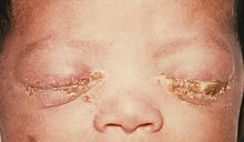
Neonatal conjunctivitis is often known as ophthalmia neonatorum. Newborns can be infected by bacteria in the birth canal. It is a disease characterized by conjunctivitis occurring in a newborn during the first month of life with clinical signs such as: erythema, and edema of the eyelids, the palpebral conjunctivae, purulent eye discharge with one or more polymorph nuclear per oil immersion field on a Gram stained conjunctival smear. Additionally, neonatal conjunctivitis is a pink eye in a newborn caused by irritation (Chemical Conjunctivitis) , a blocked tear duct, or infection.[5]
Airborne antigens may be involved in the pathogenesis of allergic conjunctivitis. Common airborne antigens, include:[6][7]
- Pollen
- Grass
- Weeds
Development of allergic conjunctivitis is result of type I hypersensitivity reactions involving the conjunctiva.
- IgE and Mast cell play an important role in these allergic inflammations.
- There is a strong association with atopic dermatitis and allergic conjunctivitis.
Combination of type I and type IV hypersensitivity reactions may be responsible in giant papillary conjunctivitis. Also prolonged mechanical irritation to the superior tarsal conjunctiva, of the upper lid, from any of a variety of foreign bodies may also be a contributing factor in giant papillary conjunctivitis.[8][9]
Keratoconjunctivitis sicca or dry eye syndrome is multifactorial disease and associated with different medical conditions such as:[10][11]
- Sjögren's syndrome
- Ocular surface disease
- Ocular allergy
- Dysfunction of the lacrimal gland with reduced tear production and Mucin deficiency
Keratoconjunctivitis sicca-associated Sjögren's syndrome may be is the result of an auto immunological sequelae which can lead to chronic inflammatory state with production of autoantibodies, such as: antinuclear antibody (ANA), rheumatoid factor (RF), fodrin (a cytoskeletal protein), the muscarinic M3 receptor, or Sjögren's syndrome-specific antibodies (anti-RO and anti-LA. Focal CD4+ T cells and B cells infiltration of the lacrimal gland can induce apoptosis in the conjunctiva and lacrimal glands and this results in dysfunction of the lacrimal gland with reduced tear production.
Superior limbic keratoconjunctivitis (SLK) is a disease characterised by inflammation of the upper palpebral and superior bulbar conjunctiva, keratinization of the superior limbus and corneal and conjunctival filaments. The exact pathogenesis of Superior limbic keratoconjunctivitis is not fully understood, however association between thyroid abnormalities (Graves ophthalmopathy) and Superior limbic keratoconjunctivitis has been reported.[12][13]
Microscopic histopathological analysis
On microscopic histopathological analysis, the following are characteristic findings of conjunctivitis:
- Mild spongiotic reaction pattern (low power view of the histology)
- Eosinophils (Allergic conjunctivitis)
- Numerous neutrophils (Bacterial conjunctivitis)
- Hyperplastic with increase numbers of plasma cell (Chronic conjunctivitis)
Gross Pathology
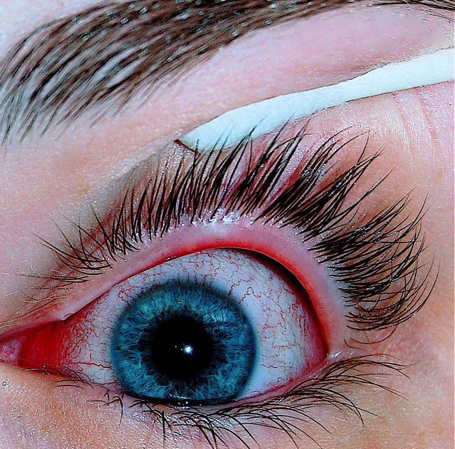
On gross pathology, the following are characteristic findings of conjunctivitis:[14]
- Conjunctival injection
- Mucopurulent or non-purulent discharge
- Pseudomembrane formation
- Chemosis
- Eyelid swelling
- Conjunctival hemorrhage (specific for Acute conjunctival hemorrhaging)
Images
The following are gross and microscopic images associated with conjunctivitis.


References
- ↑ 1.0 1.1 Azari AA, Barney NP (2013). "Conjunctivitis: a systematic review of diagnosis and treatment". JAMA. 310 (16): 1721–9. doi:10.1001/jama.2013.280318. PMC 4049531. PMID 24150468.
- ↑ 2.0 2.1 Kyei S, Koffuor GA, Ramkissoon P, Abokyi S, Owusu-Afriyie O, Wiredu EA (2015). "Possible Mechanism of Action of the Antiallergic Effect of an Aqueous Extract of Heliotropium indicum L. in Ovalbumin-Induced Allergic Conjunctivitis". J Allergy (Cairo). 2015: 245370. doi:10.1155/2015/245370. PMC 4657065. PMID 26681960.
- ↑ Everitt H, Kumar S, Little P (2003). "A qualitative study of patients' perceptions of acute infective conjunctivitis". Br J Gen Pract. 53 (486): 36–41. PMC 1314490. PMID 12564275.
- ↑ Centers for Disease Control and Prevention (2004) https://www.cdc.gov/mmwr/preview/mmwrhtml/mm5328a2.htm Accessed on June 24, 2016
- ↑ Mallika P, Asok T, Faisal H, Aziz S, Tan A, Intan G (2008). "Neonatal conjunctivitis - a review". Malays Fam Physician. 3 (2): 77–81. PMC 4170304. PMID 25606121.
- ↑ Malling HJ, Montagut A, Melac M, Patriarca G, Panzner P, Seberova E; et al. (2009). "Efficacy and safety of 5-grass pollen sublingual immunotherapy tablets in patients with different clinical profiles of allergic rhinoconjunctivitis". Clin Exp Allergy. 39 (3): 387–93. doi:10.1111/j.1365-2222.2008.03152.x. PMC 4233960. PMID 19134019.
- ↑ Kämpe M, Stålenheim G, Janson C, Stolt I, Carlson M (2007). "Systemic and local eosinophil inflammation during the birch pollen season in allergic patients with predominant rhinitis or asthma". Clin Mol Allergy. 5: 4. doi:10.1186/1476-7961-5-4. PMC 2174506. PMID 17967188.
- ↑ Donshik PC (1994). "Giant papillary conjunctivitis". Trans Am Ophthalmol Soc. 92: 687–744. PMC 1298525. PMID 7886881.
- ↑ Donshik PC, Porazinski AD (1999). "Giant papillary conjunctivitis in frequent-replacement contact lens wearers: a retrospective study". Trans Am Ophthalmol Soc. 97: 205–16, discussion 216-20. PMC 1298261. PMID 10703125.
- ↑ Zhang X, Zhao L, Deng S, Sun X, Wang N (2016). "Dry Eye Syndrome in Patients with Diabetes Mellitus: Prevalence, Etiology, and Clinical Characteristics". J Ophthalmol. 2016: 8201053. doi:10.1155/2016/8201053. PMC 4861815. PMID 27213053.
- ↑ Sivaraman KR, Jivrajka RV, Soin K, Bouchard CS, Movahedan A, Shorter E; et al. (2016). "Superior Limbic Keratoconjunctivitis-like Inflammation in Patients with Chronic Graft-Versus-Host Disease". Ocul Surf. doi:10.1016/j.jtos.2016.04.003. PMID 27179980.
- ↑ Nelson JD (1989). "Superior limbic keratoconjunctivitis (SLK)". Eye (Lond). 3 ( Pt 2): 180–9. doi:10.1038/eye.1989.26. PMID 2695351.
- ↑ Chelala E, El Rami H, Dirani A, Fakhoury H, Fadlallah A (2015). "Extensive superior limbic keratoconjunctivitis in Graves' disease: case report and mini-review of the literature". Clin Ophthalmol. 9: 467–8. doi:10.2147/OPTH.S79561. PMC 4362972. PMID 25792798.
- ↑ National Eye Institute (2015) [1] Accessed on June 24, 2016
- ↑ Image Courtesy of Joyhil09 [2]
- ↑ Image Courtesy of James Heilman [3]
- ↑ Centers for Disease Control and Prevention's Public Health Image Library[4]
- ↑ http://picasaweb.google.com/mcmumbi/USMLEIIImages