Cholera pathophysiology: Difference between revisions
No edit summary |
m (Bot: Removing from Primary care) |
||
| (51 intermediate revisions by 9 users not shown) | |||
| Line 1: | Line 1: | ||
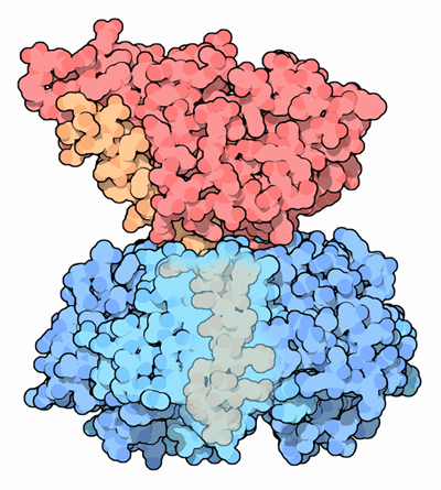
[[Image:Cholera Toxin.png|thumb|right|Cholera Toxin. The delivery region (blue) binds membrane carbohydrates to get into cells. The toxic part (red) is activated inside the cell (PDB code: 1xtc)]] | __NOTOC__ | ||
[[Image:Cholera Toxin.png|200px|thumb|right|Cholera Toxin. The delivery region (blue) binds membrane carbohydrates to get into cells. The toxic part (red) is activated inside the cell (PDB code: 1xtc)]] | |||
{{Cholera}} | {{Cholera}} | ||
{{CMG}} | {{CMG}}; '''Associate Editors-In-Chief:''' [[Priyamvada Singh|Priyamvada Singh, MBBS]], {{AA}} | ||
==Overview== | ==Overview== | ||
Cholera is mainly caused by two pathogenic serotypes of ''V. cholerae'': O1 and O139. ''V. cholerae'' is usually transmitted via the [[fecal-oral route]] to the human host. Following [[ingestion]], the ''V. cholerae'' must overcome the host defense mechanisms such as gastric acidity, intestinal inhibitory factors, and changes in temperature and [[osmolarity]]. After gaining access to [[small intestine]], ''V. cholerae'' uses [[flagella]] to propogate through the mucus layer covering the [[small intestine]] and colonizes the small intestinal cells, using toxin-coregulated pilus (TCP) to form a [[biofilm]]. [[Diarrheal]] illness in the human host is mainly caused by production of [[enterotoxin]].<ref name=Hartwell>Hartwell LH, Hood L, Goldberg ML, Reynolds AE, Silver LM, and Veres RC (2004). ''Genetics: From Genes to Genomes.'' Mc-Graw Hill, Boston: p. 551-552, 572-574 (using the turning off and turning on of [[gene expression]] to make toxin proteins in cholera bacteria as a "comprehensive example" of what is known about the mechanisms by which bacteria change the mix of proteins they manufacture to respond to the changing opportunities for surviving and thriving in different chemical environments).</ref> <ref name="pmid198781">{{cite journal| author=Cassel D, Selinger Z| title=Mechanism of adenylate cyclase activation by cholera toxin: inhibition of GTP hydrolysis at the regulatory site. | journal=Proc Natl Acad Sci U S A | year= 1977 | volume= 74 | issue= 8 | pages= 3307-11 | pmid=198781 | doi= | pmc=431542 | url=https://www.ncbi.nlm.nih.gov/entrez/eutils/elink.fcgi?dbfrom=pubmed&tool=sumsearch.org/cite&retmode=ref&cmd=prlinks&id=198781 }} </ref><ref name="pmid9841673">{{cite journal| author=Faruque SM, Albert MJ, Mekalanos JJ| title=Epidemiology, genetics, and ecology of toxigenic Vibrio cholerae. | journal=Microbiol Mol Biol Rev | year= 1998 | volume= 62 | issue= 4 | pages= 1301-14 | pmid=9841673 | doi= | pmc=98947 | url=https://www.ncbi.nlm.nih.gov/entrez/eutils/elink.fcgi?dbfrom=pubmed&tool=sumsearch.org/cite&retmode=ref&cmd=prlinks&id=9841673 }} </ref><ref name="pmid8389476">{{cite journal| author=Trucksis M, Galen JE, Michalski J, Fasano A, Kaper JB| title=Accessory cholera enterotoxin (Ace), the third toxin of a Vibrio cholerae virulence cassette. | journal=Proc Natl Acad Sci U S A | year= 1993 | volume= 90 | issue= 11 | pages= 5267-71 | pmid=8389476 | doi= | pmc=46697 | url=https://www.ncbi.nlm.nih.gov/entrez/eutils/elink.fcgi?dbfrom=pubmed&tool=sumsearch.org/cite&retmode=ref&cmd=prlinks&id=8389476 }} </ref><ref name="pmid4329549">{{cite journal| author=Hendrix TR| title=The pathophysiology of cholera. | journal=Bull N Y Acad Med | year= 1971 | volume= 47 | issue= 10 | pages= 1169-80 | pmid=4329549 | doi= | pmc=1749961 | url=https://www.ncbi.nlm.nih.gov/entrez/eutils/elink.fcgi?dbfrom=pubmed&tool=sumsearch.org/cite&retmode=ref&cmd=prlinks&id=4329549 }} </ref><ref name="pmid14407057">{{cite journal| author=JENKIN CR, ROWLEY D| title=Possible factors in the pathogenesis of cholera. | journal=Br J Exp Pathol | year= 1959 | volume= 40 | issue= | pages= 474-81 | pmid=14407057 | doi= | pmc=2082309 | url=https://www.ncbi.nlm.nih.gov/entrez/eutils/elink.fcgi?dbfrom=pubmed&tool=sumsearch.org/cite&retmode=ref&cmd=prlinks&id=14407057 }} </ref><ref name=DiRita_1991>{{cite journal |author=DiRita V, Parsot C, Jander G, Mekalanos J |title=Regulatory cascade controls virulence in Vibrio cholerae |journal=Proc Natl Acad Sci U S A |volume=88 |issue=12 |pages=5403-7 |year=1991 | url=http://www.pnas.org/cgi/reprint/88/12/5403 |id=PMID 2052618}}</ref><ref name="pmid2883655">{{cite journal| author=Taylor RK, Miller VL, Furlong DB, Mekalanos JJ| title=Use of phoA gene fusions to identify a pilus colonization factor coordinately regulated with cholera toxin. | journal=Proc Natl Acad Sci U S A | year= 1987 | volume= 84 | issue= 9 | pages= 2833-7 | pmid=2883655 | doi= | pmc=304754 | url=https://www.ncbi.nlm.nih.gov/entrez/eutils/elink.fcgi?dbfrom=pubmed&tool=sumsearch.org/cite&retmode=ref&cmd=prlinks&id=2883655 }} </ref><ref name="pmid208069">{{cite journal| author=Cassel D, Pfeuffer T| title=Mechanism of cholera toxin action: covalent modification of the guanyl nucleotide-binding protein of the adenylate cyclase system. | journal=Proc Natl Acad Sci U S A | year= 1978 | volume= 75 | issue= 6 | pages= 2669-73 | pmid=208069 | doi= | pmc=392624 | url=https://www.ncbi.nlm.nih.gov/entrez/eutils/elink.fcgi?dbfrom=pubmed&tool=sumsearch.org/cite&retmode=ref&cmd=prlinks&id=208069 }} </ref><ref name="pmid8658163">{{cite journal| author=Waldor MK, Mekalanos JJ| title=Lysogenic conversion by a filamentous phage encoding cholera toxin. | journal=Science | year= 1996 | volume= 272 | issue= 5270 | pages= 1910-4 | pmid=8658163 | doi= | pmc= | url=https://www.ncbi.nlm.nih.gov/entrez/eutils/elink.fcgi?dbfrom=pubmed&tool=sumsearch.org/cite&retmode=ref&cmd=prlinks&id=8658163 }} </ref> | |||
==Pathophysiology== | |||
Cholera is mainly caused by two pathogenic serotypes of ''V. cholerae'': O1 and O139. The pathogenesis underlying acute [[diarrheal]] illness is as follows:<ref name=Hartwell>Hartwell LH, Hood L, Goldberg ML, Reynolds AE, Silver LM, and Veres RC (2004). ''Genetics: From Genes to Genomes.'' Mc-Graw Hill, Boston: p. 551-552, 572-574 (using the turning off and turning on of [[gene expression]] to make toxin proteins in cholera bacteria as a "comprehensive example" of what is known about the mechanisms by which bacteria change the mix of proteins they manufacture to respond to the changing opportunities for surviving and thriving in different chemical environments).</ref><ref name="pmid198781">{{cite journal| author=Cassel D, Selinger Z| title=Mechanism of adenylate cyclase activation by cholera toxin: inhibition of GTP hydrolysis at the regulatory site. | journal=Proc Natl Acad Sci U S A | year= 1977 | volume= 74 | issue= 8 | pages= 3307-11 | pmid=198781 | doi= | pmc=431542 | url=https://www.ncbi.nlm.nih.gov/entrez/eutils/elink.fcgi?dbfrom=pubmed&tool=sumsearch.org/cite&retmode=ref&cmd=prlinks&id=198781 }} </ref><ref name="pmid9841673">{{cite journal| author=Faruque SM, Albert MJ, Mekalanos JJ| title=Epidemiology, genetics, and ecology of toxigenic Vibrio cholerae. | journal=Microbiol Mol Biol Rev | year= 1998 | volume= 62 | issue= 4 | pages= 1301-14 | pmid=9841673 | doi= | pmc=98947 | url=https://www.ncbi.nlm.nih.gov/entrez/eutils/elink.fcgi?dbfrom=pubmed&tool=sumsearch.org/cite&retmode=ref&cmd=prlinks&id=9841673 }} </ref><ref name="pmid8389476">{{cite journal| author=Trucksis M, Galen JE, Michalski J, Fasano A, Kaper JB| title=Accessory cholera enterotoxin (Ace), the third toxin of a Vibrio cholerae virulence cassette. | journal=Proc Natl Acad Sci U S A | year= 1993 | volume= 90 | issue= 11 | pages= 5267-71 | pmid=8389476 | doi= | pmc=46697 | url=https://www.ncbi.nlm.nih.gov/entrez/eutils/elink.fcgi?dbfrom=pubmed&tool=sumsearch.org/cite&retmode=ref&cmd=prlinks&id=8389476 }} </ref><ref name="pmid4329549">{{cite journal| author=Hendrix TR| title=The pathophysiology of cholera. | journal=Bull N Y Acad Med | year= 1971 | volume= 47 | issue= 10 | pages= 1169-80 | pmid=4329549 | doi= | pmc=1749961 | url=https://www.ncbi.nlm.nih.gov/entrez/eutils/elink.fcgi?dbfrom=pubmed&tool=sumsearch.org/cite&retmode=ref&cmd=prlinks&id=4329549 }} </ref><ref name="pmid14407057">{{cite journal| author=JENKIN CR, ROWLEY D| title=Possible factors in the pathogenesis of cholera. | journal=Br J Exp Pathol | year= 1959 | volume= 40 | issue= | pages= 474-81 | pmid=14407057 | doi= | pmc=2082309 | url=https://www.ncbi.nlm.nih.gov/entrez/eutils/elink.fcgi?dbfrom=pubmed&tool=sumsearch.org/cite&retmode=ref&cmd=prlinks&id=14407057 }} </ref><ref name=DiRita_1991>{{cite journal |author=DiRita V, Parsot C, Jander G, Mekalanos J |title=Regulatory cascade controls virulence in Vibrio cholerae |journal=Proc Natl Acad Sci U S A |volume=88 |issue=12 |pages=5403-7 |year=1991 | url=http://www.pnas.org/cgi/reprint/88/12/5403 |id=PMID 2052618}}</ref><ref name="pmid2883655">{{cite journal| author=Taylor RK, Miller VL, Furlong DB, Mekalanos JJ| title=Use of phoA gene fusions to identify a pilus colonization factor coordinately regulated with cholera toxin. | journal=Proc Natl Acad Sci U S A | year= 1987 | volume= 84 | issue= 9 | pages= 2833-7 | pmid=2883655 | doi= | pmc=304754 | url=https://www.ncbi.nlm.nih.gov/entrez/eutils/elink.fcgi?dbfrom=pubmed&tool=sumsearch.org/cite&retmode=ref&cmd=prlinks&id=2883655 }} </ref><ref name="pmid208069">{{cite journal| author=Cassel D, Pfeuffer T| title=Mechanism of cholera toxin action: covalent modification of the guanyl nucleotide-binding protein of the adenylate cyclase system. | journal=Proc Natl Acad Sci U S A | year= 1978 | volume= 75 | issue= 6 | pages= 2669-73 | pmid=208069 | doi= | pmc=392624 | url=https://www.ncbi.nlm.nih.gov/entrez/eutils/elink.fcgi?dbfrom=pubmed&tool=sumsearch.org/cite&retmode=ref&cmd=prlinks&id=208069 }} </ref><ref name="pmid8658163">{{cite journal| author=Waldor MK, Mekalanos JJ| title=Lysogenic conversion by a filamentous phage encoding cholera toxin. | journal=Science | year= 1996 | volume= 272 | issue= 5270 | pages= 1910-4 | pmid=8658163 | doi= | pmc= | url=https://www.ncbi.nlm.nih.gov/entrez/eutils/elink.fcgi?dbfrom=pubmed&tool=sumsearch.org/cite&retmode=ref&cmd=prlinks&id=8658163 }} </ref> | |||
===Transmission=== | |||
*''V. cholerae'' is usually transmitted via the [[fecal-oral route]] to the human host. | |||
*Following [[ingestion]], the ''V. cholerae'' must overcome host defense mechanisms such as gastric acidity, intestinal inhibitory factors, and changes in temperature and [[osmolarity]]. | |||
*Infective dose varies from 102-106. | |||
*The [[incubation period]] varies from a few hours to a few days. | |||
[[ | ===Colonization=== | ||
*After gaining access to [[small intestine]], ''V. cholerae'' uses [[flagella]] to propagate through the mucus layer covering [[small intestine]] and colonizes the small intestinal cells using toxin-coregulated pilus (TCP) forming a [[biofilm]]. | |||
===Enterotoxin=== | |||
*[[Diarrheal]] illness in human host is mainly caused by production of [[enterotoxin]]. | |||
*The production of [[enterotoxin]] protein in the small intestinal cells is the main mechanism responsible for causing acute [[diarrheal]] illness. | |||
*It has 5B subunits and 2A subunits. | |||
**B subunits bind the [[enterocytes]] via GM1 [[ganglioside]] receptors and cause internalization of A subunits in the cells via [[endocytosis]]. | |||
**A subunits then bind and activate the [[adenylate cyclase]] enzyme in the enterocytes, increasing the levels of [[cAMP]]. | |||
*Increased levels of [[enterotoxin]] cause activation of the [[cystic fibrosis transmembrane conductance regulator]] (CFTR), causing increased secretion of water, [[sodium]], and [[chloride]] from [[enterocytes]], which causes watery [[diarrhea]]. | |||
===Virulence factors=== | |||
The different [[virulence factors]] involved in the pathogenesis of ''[[V. cholerae]]'' involve activation of [[transcription factors]] such as ToxR, TcpP, and ToxT. Different toxins expressed by these [[transcription factors]] include: | |||
*Zona occludens toxin (zot, causes invasion by decreasing intestinal tissue resistance) | |||
*Accessory cholera toxin (ace, increases fluid secretion) | |||
*Toxin-coregulated pilus (tcpA, essential colonization factor and receptor for the CTXf phage) | |||
*NAG-specific heat-labile toxin (st) | |||
*Outer membrane porin proteins (ompU and ompT) | |||
== References == | == References == | ||
{{Reflist|2}} | {{Reflist|2}} | ||
{{WikiDoc Help Menu}} | {{WikiDoc Help Menu}} | ||
{{WikiDoc Sources}} | {{WikiDoc Sources}} | ||
[[Category:Gastroenterology]] | |||
[[Category:Emergency medicine]] | |||
[[Category:Disease]] | |||
[[Category:Up-To-Date]] | |||
[[Category:Infectious disease]] | |||
[[Category:Pediatrics]] | |||
Latest revision as of 20:55, 29 July 2020

|
Cholera Microchapters |
|
Diagnosis |
|---|
|
Treatment |
|
Case Studies |
|
Cholera pathophysiology On the Web |
|
American Roentgen Ray Society Images of Cholera pathophysiology |
|
Risk calculators and risk factors for Cholera pathophysiology |
Editor-In-Chief: C. Michael Gibson, M.S., M.D. [1]; Associate Editors-In-Chief: Priyamvada Singh, MBBS, Aysha Anwar, M.B.B.S[2]
Overview
Cholera is mainly caused by two pathogenic serotypes of V. cholerae: O1 and O139. V. cholerae is usually transmitted via the fecal-oral route to the human host. Following ingestion, the V. cholerae must overcome the host defense mechanisms such as gastric acidity, intestinal inhibitory factors, and changes in temperature and osmolarity. After gaining access to small intestine, V. cholerae uses flagella to propogate through the mucus layer covering the small intestine and colonizes the small intestinal cells, using toxin-coregulated pilus (TCP) to form a biofilm. Diarrheal illness in the human host is mainly caused by production of enterotoxin.[1] [2][3][4][5][6][7][8][9][10]
Pathophysiology
Cholera is mainly caused by two pathogenic serotypes of V. cholerae: O1 and O139. The pathogenesis underlying acute diarrheal illness is as follows:[1][2][3][4][5][6][7][8][9][10]
Transmission
- V. cholerae is usually transmitted via the fecal-oral route to the human host.
- Following ingestion, the V. cholerae must overcome host defense mechanisms such as gastric acidity, intestinal inhibitory factors, and changes in temperature and osmolarity.
- Infective dose varies from 102-106.
- The incubation period varies from a few hours to a few days.
Colonization
- After gaining access to small intestine, V. cholerae uses flagella to propagate through the mucus layer covering small intestine and colonizes the small intestinal cells using toxin-coregulated pilus (TCP) forming a biofilm.
Enterotoxin
- Diarrheal illness in human host is mainly caused by production of enterotoxin.
- The production of enterotoxin protein in the small intestinal cells is the main mechanism responsible for causing acute diarrheal illness.
- It has 5B subunits and 2A subunits.
- B subunits bind the enterocytes via GM1 ganglioside receptors and cause internalization of A subunits in the cells via endocytosis.
- A subunits then bind and activate the adenylate cyclase enzyme in the enterocytes, increasing the levels of cAMP.
- Increased levels of enterotoxin cause activation of the cystic fibrosis transmembrane conductance regulator (CFTR), causing increased secretion of water, sodium, and chloride from enterocytes, which causes watery diarrhea.
Virulence factors
The different virulence factors involved in the pathogenesis of V. cholerae involve activation of transcription factors such as ToxR, TcpP, and ToxT. Different toxins expressed by these transcription factors include:
- Zona occludens toxin (zot, causes invasion by decreasing intestinal tissue resistance)
- Accessory cholera toxin (ace, increases fluid secretion)
- Toxin-coregulated pilus (tcpA, essential colonization factor and receptor for the CTXf phage)
- NAG-specific heat-labile toxin (st)
- Outer membrane porin proteins (ompU and ompT)
References
- ↑ 1.0 1.1 Hartwell LH, Hood L, Goldberg ML, Reynolds AE, Silver LM, and Veres RC (2004). Genetics: From Genes to Genomes. Mc-Graw Hill, Boston: p. 551-552, 572-574 (using the turning off and turning on of gene expression to make toxin proteins in cholera bacteria as a "comprehensive example" of what is known about the mechanisms by which bacteria change the mix of proteins they manufacture to respond to the changing opportunities for surviving and thriving in different chemical environments).
- ↑ 2.0 2.1 Cassel D, Selinger Z (1977). "Mechanism of adenylate cyclase activation by cholera toxin: inhibition of GTP hydrolysis at the regulatory site". Proc Natl Acad Sci U S A. 74 (8): 3307–11. PMC 431542. PMID 198781.
- ↑ 3.0 3.1 Faruque SM, Albert MJ, Mekalanos JJ (1998). "Epidemiology, genetics, and ecology of toxigenic Vibrio cholerae". Microbiol Mol Biol Rev. 62 (4): 1301–14. PMC 98947. PMID 9841673.
- ↑ 4.0 4.1 Trucksis M, Galen JE, Michalski J, Fasano A, Kaper JB (1993). "Accessory cholera enterotoxin (Ace), the third toxin of a Vibrio cholerae virulence cassette". Proc Natl Acad Sci U S A. 90 (11): 5267–71. PMC 46697. PMID 8389476.
- ↑ 5.0 5.1 Hendrix TR (1971). "The pathophysiology of cholera". Bull N Y Acad Med. 47 (10): 1169–80. PMC 1749961. PMID 4329549.
- ↑ 6.0 6.1 JENKIN CR, ROWLEY D (1959). "Possible factors in the pathogenesis of cholera". Br J Exp Pathol. 40: 474–81. PMC 2082309. PMID 14407057.
- ↑ 7.0 7.1 DiRita V, Parsot C, Jander G, Mekalanos J (1991). "Regulatory cascade controls virulence in Vibrio cholerae". Proc Natl Acad Sci U S A. 88 (12): 5403–7. PMID 2052618.
- ↑ 8.0 8.1 Taylor RK, Miller VL, Furlong DB, Mekalanos JJ (1987). "Use of phoA gene fusions to identify a pilus colonization factor coordinately regulated with cholera toxin". Proc Natl Acad Sci U S A. 84 (9): 2833–7. PMC 304754. PMID 2883655.
- ↑ 9.0 9.1 Cassel D, Pfeuffer T (1978). "Mechanism of cholera toxin action: covalent modification of the guanyl nucleotide-binding protein of the adenylate cyclase system". Proc Natl Acad Sci U S A. 75 (6): 2669–73. PMC 392624. PMID 208069.
- ↑ 10.0 10.1 Waldor MK, Mekalanos JJ (1996). "Lysogenic conversion by a filamentous phage encoding cholera toxin". Science. 272 (5270): 1910–4. PMID 8658163.