Chancroid physical examination
|
Chancroid Microchapters |
|
Diagnosis |
|---|
|
Treatment |
|
Case Studies |
|
Chancroid physical examination On the Web |
|
American Roentgen Ray Society Images of Chancroid physical examination |
|
calculators and risk factors for Chancroid physical examination |
Editor-In-Chief: C. Michael Gibson, M.S., M.D. [1]; Associate Editor(s)-in-Chief: Yazan Daaboul, M.D. Nate Michalak, B.A. Serge Korjian M.D.
Overview
Physical Examination
Vital Signs
Typically normal
Skin
A patient may present with either of the following types of lesions on the genitals, depending on the stage of infection:[1][2]
Ulcer characteristics:[3]
- Ranges in size from 3 to 50 mm (1/8 to 2 inches) in diameter
- Painful
- Soft, nonindurated
- Irregular border
- Sharp margins
- Grey/yellow exudate
- Males typically have a single ulcer
- Females typically have multiplee ulcers
Common locations in males
- Foreskin (prepuce) (most common)
- Groove behind the head of the penis (coronal sulcus)
- Shaft of the penis
- Head of the penis (glans penis)
- Opening of the penis (urethral meatus)
- Scrotum (least common)
Common locations in females
- labia majora (most common). "Kissing ulcers" may develop, defined as ulcers that occur on opposing surfaces of the labia.
- labia minora
- Perineal area
- Inner thighs (least common)
The initial ulcer may be mistaken as a "hard" chancre, the typical sore of primary syphilis, as opposed to the "soft chancre" of chancroid.
Approximately one third of the infected individuals will develop enlargements of the inguinal lymph nodes, the nodes located in the fold between the leg and the lower abdomen.
Half of those who develop swelling of the inguinal lymph nodes will progress to a point where the nodes rupture through the skin producing draining abscesses. The swollen lymph nodes and abscesses are often referred to as buboes.
Skin
Genital
-
![Chancroid. Adapted from Dermatology Atlas.[4]](/images/6/6f/Chancroid01.jpg)
Chancroid. Adapted from Dermatology Atlas.[4]
-
![Chancroid. Adapted from Dermatology Atlas.[4]](/images/c/c1/Chancroid02.jpg)
Chancroid. Adapted from Dermatology Atlas.[4]
-
![Chancroid. Adapted from Dermatology Atlas.[4]](/images/1/1a/Chancroid03.jpg)
Chancroid. Adapted from Dermatology Atlas.[4]
-
![Chancroid. Adapted from Dermatology Atlas.[4]](/images/7/70/Chancroid04.jpg)
Chancroid. Adapted from Dermatology Atlas.[4]
-
![Chancroid. Adapted from Dermatology Atlas.[4]](/images/2/22/Chancroid05.jpg)
Chancroid. Adapted from Dermatology Atlas.[4]
-
![Chancroid. Adapted from Dermatology Atlas.[4]](/images/d/d6/Chancroid06.jpg)
Chancroid. Adapted from Dermatology Atlas.[4]
-
![Chancroid. Adapted from Dermatology Atlas.[4]](/images/1/19/Chancroid07.jpg)
Chancroid. Adapted from Dermatology Atlas.[4]
-
![Chancroid. Adapted from Dermatology Atlas.[4]](/images/6/60/Chancroid08.jpg)
Chancroid. Adapted from Dermatology Atlas.[4]
-
![Chancroid. Adapted from Dermatology Atlas.[4]](/images/4/4b/Chancroid09.jpg)
Chancroid. Adapted from Dermatology Atlas.[4]
-
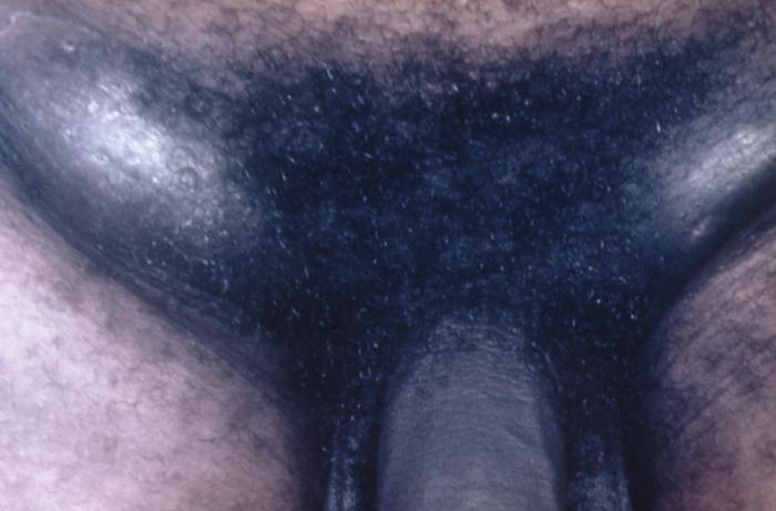
Chancroid infection has spread to the inguinal lymph nodes, which have enlarged forming buboes.
-
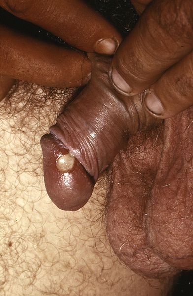
Penile chancroid lesion.
![Chancroid. Adapted from Dermatology Atlas.[4]](/index.php/File:Chancroid01.jpg)
![Chancroid. Adapted from Dermatology Atlas.[4]](/index.php/File:Chancroid02.jpg)
![Chancroid. Adapted from Dermatology Atlas.[4]](/index.php/File:Chancroid03.jpg)
![Chancroid. Adapted from Dermatology Atlas.[4]](/index.php/File:Chancroid04.jpg)
![Chancroid. Adapted from Dermatology Atlas.[4]](/index.php/File:Chancroid05.jpg)
![Chancroid. Adapted from Dermatology Atlas.[4]](/index.php/File:Chancroid06.jpg)
![Chancroid. Adapted from Dermatology Atlas.[4]](/index.php/File:Chancroid07.jpg)
![Chancroid. Adapted from Dermatology Atlas.[4]](/index.php/File:Chancroid08.jpg)
![Chancroid. Adapted from Dermatology Atlas.[4]](/index.php/File:Chancroid09.jpg)


References
- ↑ Chancroid. UpToDate (September 25, 2015). http://www.uptodate.com/contents/chancroid#H3 Accessed January 19, 2016.
- ↑ Spinola, S. M. (2002). "Immunopathogenesis of Haemophilus ducreyi Infection (Chancroid)". Infection and Immunity. 70 (4): 1667–1676. doi:10.1128/IAI.70.4.1667-1676.2002. ISSN 0019-9567.
- ↑ Chancroid. Wikipedia (July 16, 2015). https://en.wikipedia.org/wiki/Chancroid Accessed January 15, 2016.
- ↑ 4.0 4.1 4.2 4.3 4.4 4.5 4.6 4.7 4.8 "Dermatology Atlas".