Cardiac tamponade pathophysiology: Difference between revisions
Varun Kumar (talk | contribs) No edit summary |
No edit summary |
||
| Line 1: | Line 1: | ||
==Pathophysiology== | ==Pathophysiology== | ||
The outer pericardium is made of fibrous tissue <ref>Thibodeau, G.A., Patton, K.T. (2000). Anatomy & Physiology. Missouri: Mosby ISBN 9780323010962 </ref> which does not easily stretch, and so once fluid begins to enter the pericardial space, pressure starts to increase <ref>Mattson Porth, C. (Ed.) (2005) (7th Ed.) Pathophysiology: Concepts of Altered Health States. Philadelphia : Lippincott Williams & Wilkins ISBN 978-0781749886 </ref>. | The outer pericardium is made of fibrous tissue <ref>Thibodeau, G.A., Patton, K.T. (2000). Anatomy & Physiology. Missouri: Mosby ISBN 9780323010962 </ref> which does not easily stretch, and so once fluid begins to enter the pericardial space, pressure starts to increase <ref>Mattson Porth, C. (Ed.) (2005) (7th Ed.) Pathophysiology: Concepts of Altered Health States. Philadelphia : Lippincott Williams & Wilkins ISBN 978-0781749886 </ref>. | ||
Revision as of 18:17, 26 March 2012
Pathophysiology
The outer pericardium is made of fibrous tissue [1] which does not easily stretch, and so once fluid begins to enter the pericardial space, pressure starts to increase [2].
Ordinarily drainage from the pericardium occurs via the thoracic duct and the right lymphatic duct into the right pleural space. In the absence of disease, the normal pericardium contains only 20-50 cc of serous fluid due to ultrafiltration from the blood. Up to about 75 cc can accumulate acutely in the pericardium without hemodynamic compromise. Much greater amounts of fluid can accumulate chronically over a prolonged period of time as the pericardial sac stretches slowly to accommodate the fluid without hemodynamic compromise. However, if the volume of the fluid accumulation is too rapid and or large, then hemodynamic compromise can occur with a rise in pericardial pressure. This in turn reduces stroke volume and eventually cardiac output.
If fluid continues to accumulate, then with each successive diastole, less and less blood enters the ventricles, as the increasing pressure presses on the heart and forces the septum to bend into the left ventricle, leading to decreased stroke volume [3]. This causes obstructive shock to develop, and if left untreated then Cardiac arrest may occur (in which case the presenting rhythm is likely to be Pulseless electrical activity)
Below is a video demonstrating hemorrhagic effusion leading to cardiac tamponade. <youtube v=QwgfuDegC5Y/>
Autopsy Studies of Cardiac Tamponade
-
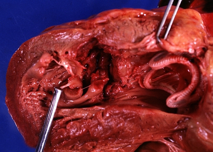
HEART: Myocardial Rupture Following Mitral Valve Replacement: Gross, an excellent example with valve and obviously ruptured heart wall.
-
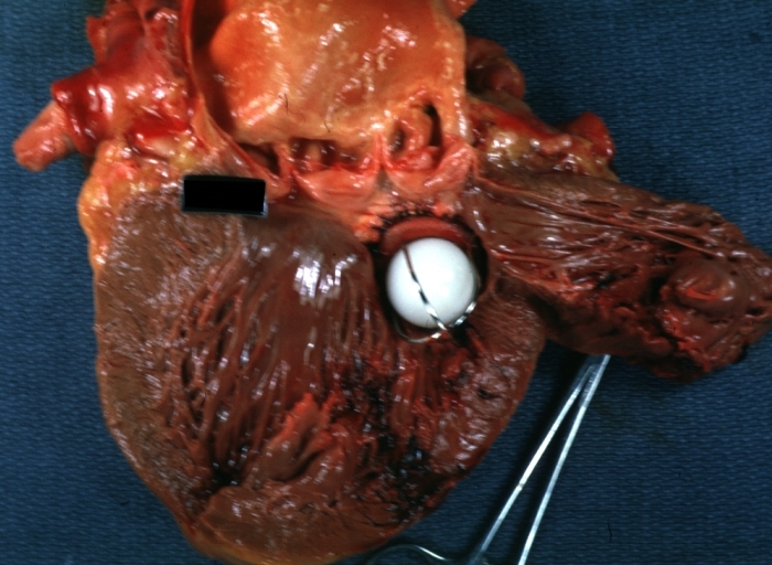
HEART: Myocardial Rupture Following Mitral Valve Replacement: Gross, natural color view from within left ventricle caged plastic ball with steel complete struts


-
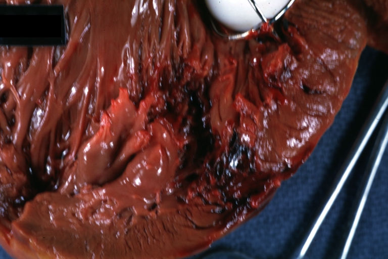
HEART: Myocardial Rupture Following Mitral Valve Replacement: Gross, natural color portion of caged plastic ball prosthesis shown with close-up of ruptured left ventricular wall
-
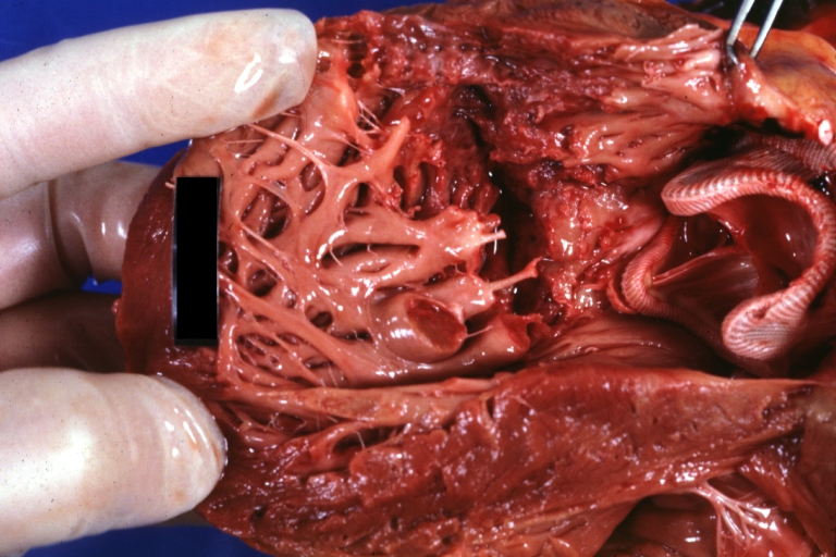
HEART: Myocardial Rupture Following Mitral Valve Replacement: Gross, natural color close-up view of torn myocardium and bottom of hetero or homograft valve


-
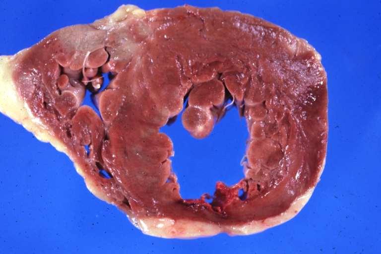
HEART: Infarct; Acute, Ruptured: Gross, natural color horizontal section ventricle anterior infarct not easily appreciated with thinning of myocardium
-
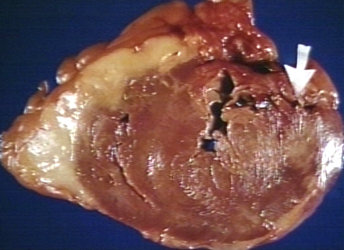
HEART: Rupture after myocardial infarction.


References
- ↑ Thibodeau, G.A., Patton, K.T. (2000). Anatomy & Physiology. Missouri: Mosby ISBN 9780323010962
- ↑ Mattson Porth, C. (Ed.) (2005) (7th Ed.) Pathophysiology: Concepts of Altered Health States. Philadelphia : Lippincott Williams & Wilkins ISBN 978-0781749886
- ↑ Mattson Porth, C. (Ed.) (2005) (7th Ed.) Pathophysiology: Concepts of Altered Health States. Philadelphia : Lippincott Williams & Wilkins ISBN 978-0781749886