Thoracic duct
Editor-In-Chief: C. Michael Gibson, M.S., M.D. [1]
In human anatomy, the thoracic duct is an important part of the lymphatic system—it is the largest lymphatic vessel in the body.
It collects most of the lymph in the body (except that from the right arm and the right side of the chest, neck and head, which is collected by the right lymphatic duct) and drains into the systemic (blood) circulation at the left subclavian vein.
Location and direction of flow
In adults, the thoracic duct is typically 38-45cm in length and an average diameter of about 5mm. It usually starts from the level of the second lumbar vertebra and extends to the root of the neck.
It originates in the abdomen from the confluence of the right and left lumbar trunk and the intestinal trunk, forming a significant pathway upward called the cisterna chyli.
It extends vertically in the chest and curves posteriorly to the left carotid artery and left jugular vein at the C7 vertebral level to empty into the junction of the left subclavian vein and left jugular vein, below the clavicle, near the shoulders.
It traverses the diaphragm at the aortic aperture and ascends the posterior mediastinum between the descending thoracic aorta (to its left) and the azygos vein (to its right).
Volume, mechanism, and direction of flow
In adults, the thoracic duct transports up to 4 L of lymph per day.
The lymph transport in the thoracic duct is mainly caused by the action of breathing, aided by the duct's smooth muscle and by internal one way valves which prevent the lymph from flowing back down again.
There are also two valves at the junction of the duct with the left subclavian vein, to prevent the flow of venous blood into the duct.
Clinical significance
When the thoracic duct is blocked or damaged a large amount of lymph can quickly accumulate in the pleural cavity, this situation is called chylothorax.
The first sign of a malignancy (especially an intraabdominal one) may be an enlarged Virchow's node, a lymph node in the left supraclavicular area, in the vicinity where the thoracic duct empties into the left subclavian vein.
Nomenclature
It is also known under various other names including the alimentary duct, chyliferous duct, duct of Pecquet, the left lymphatic duct and Van Hoorne's canal. [1]
Additional images
-
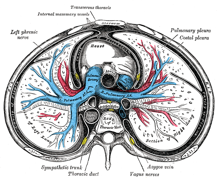 Transverse section of thorax, showing relations of pulmonary artery.
 -
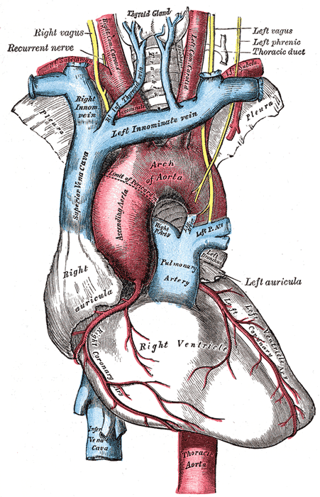 The arch of the aorta, and its branches.
 -
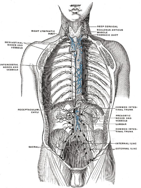 Deep lymph nodes and vessels of the thorax and abdomen (diagrammatic).
 -
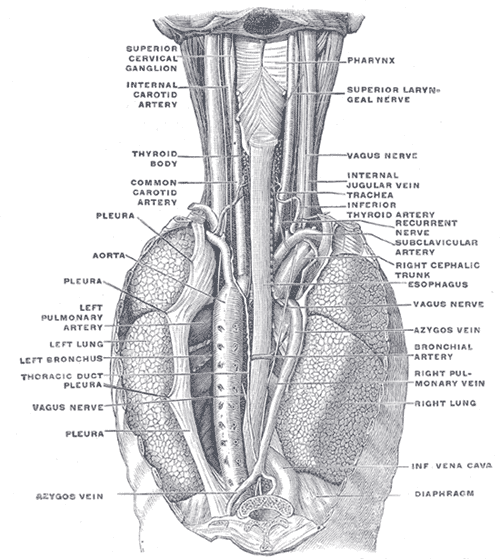 The position and relation of the esophagus in the cervical region and in the posterior mediastinum. Seen from behind.


See also
References
External links
- Template:MUNAnatomy
- Template:SUNYAnatomyFigs - "The thoracic duct and azygos venous network."
- Template:SUNYAnatomyImage
- Template:DartmouthHumanAnatomy
- Diagram at anatomyatlases.org