Atrial fibrillation cardiac MRI: Difference between revisions
No edit summary |
m (Bot: Adding CME Category::Cardiology) |
||
| Line 30: | Line 30: | ||
{{WH}} | {{WH}} | ||
{{WS}} | {{WS}} | ||
[[CME Category::Cardiology]] | |||
[[Category:Electrophysiology]] | [[Category:Electrophysiology]] | ||
Revision as of 00:56, 15 March 2016
| Resident Survival Guide |
| File:Critical Pathways.gif |
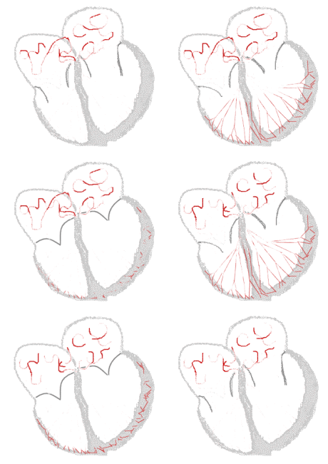
Sinus rhythm  |
Atrial fibrillation  |
|
Atrial Fibrillation Microchapters | |
|
Special Groups | |
|---|---|
|
Diagnosis | |
|
Treatment | |
|
Cardioversion | |
|
Anticoagulation | |
|
Surgery | |
|
Case Studies | |
|
Atrial fibrillation cardiac MRI On the Web | |
|
Directions to Hospitals Treating Atrial fibrillation cardiac MRI | |
|
Risk calculators and risk factors for Atrial fibrillation cardiac MRI | |
Please help WikiDoc by adding content here. It's easy! Click here to learn about editing. Editor-In-Chief: C. Michael Gibson, M.S., M.D. [1]
Overview
Cardiac magnetic resonance imaging may be used to assess the structure and the function of the atria in patients with atrial fibrillation. Further studies are needed to determine whether CMR is useful for detecting atrial thrombi in persons with atrial fibrillation.
ACCF/ACR/AHA/NASCI/SCMR 2010 Expert Consensus Document on Cardiovascular Magnetic Resonance[1] (DO NOT EDIT)
| “ |
CMR may be used for assessing left atrial structure and function in patients with atrial fibrillation. The writing committee recognizes that evolving techniques utilizing LGE may have high utility for identifying evidence of fibrotic tissue within the atrial wall or an adjoining structure. Standardization of protocols and further studies are needed to determine if CMR provides a reliable effective method for detecting thrombi in the left atrial appendage in patients with atrial fibrillation. CMR is recommended for identifying pulmonary vein anatomy prior to or after electrophysiology procedures without need for patient exposure to ionizing radiation. |
” |
References
- ↑ American College of Cardiology Foundation Task Force on Expert Consensus Documents. Hundley WG, Bluemke DA, Finn JP, Flamm SD, Fogel MA; et al. (2010). "ACCF/ACR/AHA/NASCI/SCMR 2010 expert consensus document on cardiovascular magnetic resonance: a report of the American College of Cardiology Foundation Task Force on Expert Consensus Documents". Circulation. 121 (22): 2462–508. doi:10.1161/CIR.0b013e3181d44a8f. PMC 3034132. PMID 20479157.