|
|
| (19 intermediate revisions by 3 users not shown) |
| Line 1: |
Line 1: |
| __NOTOC__ | | __NOTOC__ |
| {{Astrocytoma}} | | {{Astrocytoma}} |
| {{CMG}}; {{AE}} {{Ammu}} | | {{CMG}}; {{AE}} {{Fs}} |
| ==Overview== | | ==Overview== |
| On [[MRI]] of [[head]], astrocytoma is characterized by non-enhancing [[isointense]] to [[hypointense]] lesions compared to [[white matter]].
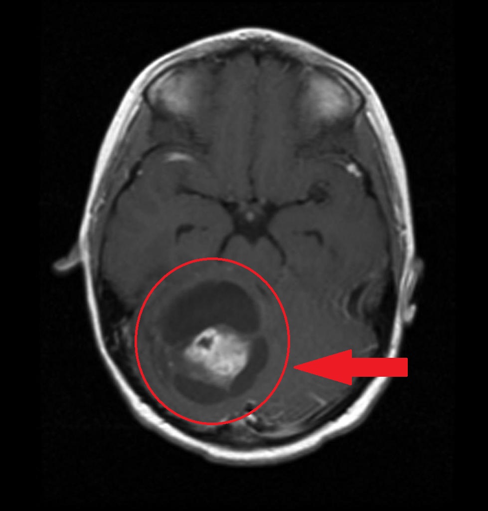
| | Findings on MRI suggestive of astrocytoma in [[Low grade astrocytoma]] ([[Pilocytic astrocytoma|pilocytic]] and [[diffuse astrocytoma]]) include Decreased resonance in comparison to surrounding brain tissue in T1 and Increased resonance in comparison to surrounding brain tissue in T2. In [[anaplastic astrocytoma]] we have Hypointense T1, Hyperintense T2 and some contrast enhancement and [[edema]]. In [[Glioblastoma multiforme|glioblastoma multiform]] we have irregular ring-nodular enhancing lesions and central [[necrosis]] surrounding [[vasogenic edema]]. |
| | |
| ==MRI== | | ==MRI== |
| ===Low grade infiltrative astrocytoma<ref name=Radiopaedia2015>{{cite web | title = Low grade infiltrative astrocytoma [Dr Bruno Di Muzio and Dr Frank Gaillard]| url = http://radiopaedia.org/articles/low-grade-infiltrative-astrocytoma }}</ref>=== | | [[MRI]] may be helpful in the diagnosis of astrocytoma. Findings on MRI suggestive of astrocytoma include:<ref name="pmid17964028">{{cite journal |vauthors=Sathornsumetee S, Rich JN, Reardon DA |title=Diagnosis and treatment of high-grade astrocytoma |journal=Neurol Clin |volume=25 |issue=4 |pages=1111–39, x |date=November 2007 |pmid=17964028 |doi=10.1016/j.ncl.2007.07.004 |url=}}</ref><ref name="pmid22819718">{{cite journal |vauthors=Pedersen CL, Romner B |title=Current treatment of low grade astrocytoma: a review |journal=Clin Neurol Neurosurg |volume=115 |issue=1 |pages=1–8 |date=January 2013 |pmid=22819718 |doi=10.1016/j.clineuro.2012.07.002 |url=}}</ref> |
| * [[MRI]] is the modality of choice for characterising these [[lesion]]s, and in the case of smaller [[tumor]]s, they may be subtle and difficult to see on [[CT]], especially as they tend not to enhance. | | * [[Low grade astrocytoma]] ([[Pilocytic astrocytoma|pilocytic]] and [[diffuse astrocytoma]]) |
| * Reported signal characteristics include: | | ** T1: Decreased resonance in comparison to surrounding brain tissue |
| * T1 | | ** T2: Increased resonance in comparison to surrounding brain tissue |
| :* [[Isointense]] to [[hypointense]] compared to [[white matter]] | | * [[High grade astrocytoma]] |
| :* Usually confined to the [[white matter]]s and causes expansion of the adjacent [[cortex]]
| | ** [[Anaplastic astrocytoma|Anaplastic astrocytomas]] |
| * T2/FLAIR | | *** Hypointense T1 |
| :* [[Mass]]-like [[hyperintense]] signals
| | *** Hyperintense T2 |
| :* Always follow the [[white matter]] distribution and cause expansion of the surrounding [[cortex]]
| | *** There might be some contrast enhancement and [[edema]] |
| :* [[Cortex]] can also, be involved in late cases in comparison to the [[oligodendroglioma]] which is a cortical based [[tumor]] from the start
| | ** [[Glioblastoma multiforme|Glioblastoma multiform]] |
| :* The "micro-cystic changes" along the lines of spread of the infiltrative astrocytoma is a very unique behavior for the infilterative astrocytoma, however, it is only appreciated in a few number of cases
| | *** Irregular ring-nodular enhancing lesions |
| :* High T2 signal is NOT related to cellularity or cellular [[atypia]], but rather [[edema]], [[demyelination]] and other degenerative changes
| | *** Central [[necrosis]] |
| * DWI
| | *** Surrounding [[vasogenic edema]] |
| :* No restricted diffusion
| |
| :* Increased diffusibility is the key to differentiate the diffuse astrocytoma from the acute [[ischemia]]
| |
| * T1 C+ (Gd) | |
| :* No enhancement is often the rule but small ill defined area of enhancement is not rare (as in path proven case 1); however, when enhancement is seen it should be considered as a warning sign for a progression to a higher grade
| |
| * [[MR spectroscopy]] | |
| :* Typical [[tumor]] will show elevated [[choline]] peak, low NAA peak, elevated [[choline:creatine]] ratio
| |
| :* There is lack of the [[lactate]] peak seen at 1:33
| |
| :* The [[lactate]] peak represents the [[necrosis]] seen in aggressive mostly WHO grade IV [[tumor]]s
| |
| :* [[MR perfusion]]: no elevation of rCBV
| |
|
| |
|
| ====Fibrillary Astrocytoma<ref name=Radiopaedia2-2015>{{cite web | title = Fibrillary Astrocytoma [Dr Frank Gaillard]| url = http://radiopaedia.org/articles/fibrillary-astrocytoma }}</ref>==== | | ===== Pilocytic astrocytoma ===== |
| * T1: [[isointense]] to [[hypointense]] compared to [[white matter]]
| |
| * T2: [[hyperintense]]
| |
| * T1 C+ (Gd): usually little or no enhancement
| |
| * MR [[spectroscopy]]: elevated [[choline]]:[[creatine]] ratio
| |
| * MR [[perfusion]]: no elevation of rCBV
| |
|
| |
|
| ====Protoplasmic Astrocytoma<ref name=Radiopaedia3-2015>{{cite web | title = Protoplasmic astrocytoma [Dr Yuranga Weerakkody and Dr Frank Gaillard]| url = http://radiopaedia.org/articles/protoplasmic-astrocytoma }}</ref>====
| | [[File:AAstrocytoma.jpg|400px|none|thumb|Source Radiopedia by A.Prof Frank Gaillard <ref>Case courtesy of A.Prof Frank Gaillard, <a href="https://radiopaedia.org/">Radiopaedia.org</a>. From the case <a href="https://radiopaedia.org/cases/8474">rID: 8474</a></ref>]] |
| * These [[tumor]]s have fairly characteristic appearances<ref name="pmid20644924">{{cite journal| author=Tay KL, Tsui A, Phal PM, Drummond KJ, Tress BM| title=MR imaging characteristics of protoplasmic astrocytomas. | journal=Neuroradiology | year= 2011 | volume= 53 | issue= 6 | pages= 405-11 | pmid=20644924 | doi=10.1007/s00234-010-0741-2 | pmc= | url=http://www.ncbi.nlm.nih.gov/entrez/eutils/elink.fcgi?dbfrom=pubmed&tool=sumsearch.org/cite&retmode=ref&cmd=prlinks&id=20644924 }} </ref>:
| |
| :* T1: [[hypointense]] compared to [[white matter]]
| |
| :* T2: strikingly [[hyperintense]]
| |
| :* FLAIR: large areas of T2 [[hyperintensity]] suppress on FLAIR (these are not macrocystic but rather represent the areas with abundant microcystic change)
| |
| :* T1 C+ (Gd): usually little or no enhancement
| |
| :* [[MR spectroscopy]]: elevated [[choline]]:[[creatine]] ratio
| |
| :* MR [[perfusion]]: there is reduced rCBV
| |
| * The key features which should prompt a protoplasmic astrocytoma being raised as the favored [[diagnosis]] are: A) prominent involvement of [[cortex]] B) large portions of the [[tumor]] demonstrating high T2 signal which suppresses on FLAIR.
| |
|
| |
|
| ===Anaplastic astrocytomas<ref name=Radiopaedia 2015 Anaplastic astrocytoma>{{cite web | title = Anaplastic astrocytoma [Dr Bruno Di Muzio and Dr Frank Gaillard]| url = http://radiopaedia.org/articles/anaplastic-astrocytoma }}</ref>=== | | ===== Diffuse astrocytoma ===== |
| * Anaplastic astrocytomas appear similar to low grade astrocytomas but are more variable in appearance and a single [[tumor]] demonstrates more [[heterogeneity]].
| |
| * The key to distinguishing anaplastic astrocytomas from low grade [[tumor]]s is the presence of enhancement which should be absent in the latter (although one should note that variants, especially gemistocytic astrocytomas, can demonstrate enhancement). The pattern of enhancement is very variable 1.
| |
| * Unlike [[glioblastoma]]s, anaplastic astrocytomas lack frank [[necrosis]], and as such central non-enhancing [[fluid]] intensity regions should be absent 1.
| |
| :* T1: [[hypointense]] compared to [[white matter]]
| |
| :* T2: generally [[hyperintense]] but can be heterogeneous if [[calcification]] of [[blood]] present
| |
| :* T1 C+ (Gd)
| |
| ::* Very variable but usually at least some enhancement present
| |
| ::* Presence of [[ring]] enhancement suggests central [[necrosis]] and thus [[glioblastoma]] rather than anaplastic astrocytoma
| |
| * [[MR spectroscopy]]
| |
| :* Increased [[choline]]: [[creatine]] ratio
| |
| :* NAA preserved or mildly depressed
| |
| :* No significant [[lactate]]
| |
| :* Intermediate levels of Myo-[[inositol]] (lower than low [[grade]], higher than [[glioblastoma]]
| |
| :* [[MR perfusion]]: elevated cerebral [[blood]] volume
| |
|
| |
|
| ===Pilocytic astrocytoma<ref name=Radiopaedia 2015 Pilocytic astrocytomas >{{cite web | title = Pilocytic astrocytomas [Dr Bruno Di Muzio and Dr Frank Gaillard]| url = http://radiopaedia.org/articles/pilocytic-astrocytoma }}</ref>===
| | [[File:Diffuse-astrocytoma-nos-44.jpg|400px|none|thumb|Source Radiopedia by Dr Bruno Di Muzio <ref>Case courtesy of Dr Bruno Di Muzio, <a href="https://radiopaedia.org/">Radiopaedia.org</a>. From the case <a href="https://radiopaedia.org/cases/41396">rID: 41396</a></ref>]] |
| * General
| |
| :* Pilocytic astrocytomas range in appearance | |
| :* Large cystic component with a brightly enhancing mural [[nodule]]: 67% | |
| :* Non enhancing [[cyst]] wall: 21%
| |
| :* Enhancing [[cyst]] wall: 46%
| |
| :* Heterogeneous, mixed solid and multiple [[cyst]]s and central [[necrosis]]: 16%
| |
| :* Completely solid: 17%
| |
| :* Enhancement is almost invariably present (~95%). Up to 20% may demonstrate some [[calcification]]. [[Hemorrhage]] is a rare complication.
| |
| * Signal characteristics include
| |
| :* T1: iso to [[hypointense]] solid component compared to adjacent [[brain]]
| |
| :* T2: [[hyperintense]] solid component compared to adjacent [[brain]]
| |
|
| |
|
| ===Pilomyxoid Astrocytomas<ref name=Radiopaedia 2015 Pilomyxoid astrocytoma>{{cite web | title = Pilomyxoid astrocytoma [Dr Bruno Di Muzio and Dr Imran Jindani]| url = http://radiopaedia.org/articles/pilomyxoid-astrocytoma }}</ref>=== | | === Anaplastic astrocytoma === |
| * Reported signal characteristics include
| |
| :* T1: [[isointense]]
| |
| :* T2: usually [[hyperintense]]
| |
| :* T1 C+ (Gd): common and is usually in the solid component, but can be also peripheric
| |
| {|
| |
| | valign=top |
| |
| [[File:Pilocystic 01.jpg|thumb|center|200 px|Stereotactic MRI brain showed recurrent postoperative brain stem cystic pilocytic astrocytoma.<SMALL><SMALL>''[https://en.wikipedia.org/wiki/Pilocytic_astrocytoma#/media/File:Pilocytic.jpg]''<ref name="Wikipedia">{{Cite web | title = Wikipedia| url = https://en.wikipedia.org/wiki/Pilocytic_astrocytoma#/media/File:Pilocytic.jpg}}</ref></SMALL></SMALL>]]
| |
| |style="width: 40px"|
| |
| [[File:Pilocystic 04.jpg|thumb|center|200 px|Sagittal T1-weighted MRI showing a well circumscribed hypointense mass in the tectum presumably a tectal plate glioma. These lesions are a distinct subset of pilocytic astrocytomas which present with hydrocephalus in 6 to 10 year olds and are rarely progressive lesions, when imaging is characteristic, biopsy is usually not performed because of the risks to adjacent structures, often shunting is the only treatment required.<SMALL><SMALL>''[https://en.wikipedia.org/wiki/Pilocytic_astrocytoma#/media/File:Pilocytic.jpg]''<ref name="Wikipedia">{{Cite web | title = Wikipedia| url = https://en.wikipedia.org/wiki/Pilocytic_astrocytoma#/media/File:Pilocytic.jpg}}</ref></SMALL></SMALL>]]
| |
| |style="width: 40px"|
| |
| | [[File:Pilocystic 05.jpg|thumb|center|200 px|T2-weighted coronal MRI in the same patient showing the hyper- intense lesion to originate just to the right of midline with deviation and compression and obstruction of the aqueduct with resultant dilation of the lateral ventricles<SMALL><SMALL>''[https://en.wikipedia.org/wiki/Pilocytic_astrocytoma#/media/File:Pilocytic.jpg]''<ref name="Wikipedia">{{Cite web | title = Wikipedia| url = https://en.wikipedia.org/wiki/Pilocytic_astrocytoma#/media/File:Pilocytic.jpg}}</ref></SMALL></SMALL>]]
| |
| |style="width: 40px"|
| |
| |[[File:Pilocystic 06.jpg|thumb|center|200 px|Axial FLAIR MRI in the same patient showing the lesion to be hyperintense, note the suppression of the CSF in the ventricular system and subarachnoid space by the FLAIR technique<SMALL><SMALL>''[https://en.wikipedia.org/wiki/Pilocytic_astrocytoma#/media/File:Pilocytic.jpg]''<ref name="Wikipedia">{{Cite web | title = Wikipedia| url = https://en.wikipedia.org/wiki/Pilocytic_astrocytoma#/media/File:Pilocytic.jpg}}</ref></SMALL></SMALL>]]
| |
| | valign=top |
| |
| [[File:Pilocystic 07.jpg|thumb|center|200 px|T1-weighted coronal MRI image post contrast showing heterogeneous contrast enhancement within the presumed tectal plate glioma <SMALL><SMALL>''[https://en.wikipedia.org/wiki/Pilocytic_astrocytoma#/media/File:Pilocytic.jpg]''<ref name="Wikipedia">{{Cite web | title = Wikipedia| url = https://en.wikipedia.org/wiki/Pilocytic_astrocytoma#/media/File:Pilocytic.jpg}}</ref></SMALL></SMALL>]]
| |
| |}
| |
|
| |
|
| ===Subependymal Giant Cell Astrocytoma<ref name=Radiopaedia052015>{{cite web | title = Subependymal giant cell astrocytoma [Dr Bruno Di Muzio and Dr Jeremy Jones]| url http://radiopaedia.org/articles/subependymal-giant-cell-astrocytoma }}</ref>===
| | [[File:Anaplastic-astrocytoma-22.jpg|400px|none|thumb|Source Radiopedia by Dr Bruno Di Muzio <ref>Case courtesy of Dr Bruno Di Muzio, <a href="https://radiopaedia.org/">Radiopaedia.org</a>. From the case <a href="https://radiopaedia.org/cases/39124">rID: 39124</a></ref>]] |
| * T1: heterogenous and hypo- to [[isointense]] to [[grey matter]]
| |
| * T2: [[heterogenous]] and [[hyperintense]] to [[grey matter]]; calcified components can be [[hypointense]]
| |
| * T1 C+ (Gd): can show marked enhancement
| |
| {|
| |
| | valign=top |
| |
| [[File:Astrocytoma 01.jpg|thumb|center|200 px|https://en.wikipedia.org/wiki/Subependymal_giant_cell_astrocytoma]]
| |
| |style="width: 300px"|
| |
| | [[File:Astrocytoma 02.jpg|thumb|center|300 px|https://en.wikipedia.org/wiki/Subependymal_giant_cell_astrocytoma]]
| |
| |}
| |
|
| |
|
| ===Pleomorphic xanthoastrocytomas (PXA)<ref name=Radiopaedia062015>{{cite web | title = Pleomorphic xanthoastrocytomas [Dr Bruno Di Muzio and Dr Frank Gaillard]| url = http://radiopaedia.org/articles/pleomorphic-xanthoastrocytoma }}</ref> ===
| |
| * T1
| |
| :* Solid component iso to [[hypointense]] c.f. [[grey matter]]
| |
| :* Cystic component low signal
| |
| :* Leptomeningeal involvement seen in over 70% of cases 2
| |
| * T1 C+ (Gd)
| |
| :* Solid component usually enhances vividly
| |
| * T2
| |
| :* Solid component iso to [[hyperintense]] c.f. [[grey matter]]
| |
| :* Cystic component high signal
| |
| :* Little surrounding vasogenic [[edema]]
| |
|
| |
|
| ===Oligoastrocytomas<ref name=Radiopaedia072015>{{cite web | title = Oligoastrocytomas [Dr Bruno Di Muzio and Dr Frank Gaillard]| url = http://radiopaedia.org/articles/oligoastrocytoma }}</ref>=== | | === Glioblastoma multiform === |
| * T1: usually [[hypointense]]
| |
| * T2: usually [[hyperintense]]
| |
| * T1 C+ (Gd): usually non-enhancing lesions
| |
|
| |
|
| ===Spinal astrocytoma<ref name=Radiopaedia112015>{{cite web | title = Spinalastrocytomas [Dr Bruno Di Muzio and Dr Frank Gaillard]| url = http://radiopaedia.org/articles/spinal-astrocytoma }}</ref>===
| | [[File:Glioblastoma-nos-butterfly-morphologyyy.jpg|400px|none|thumb|Source Radiopedia by A.Prof Frank Gaillard <ref>Case courtesy of A.Prof Frank Gaillard, <a href="https://radiopaedia.org/">Radiopaedia.org</a>. From the case <a href="https://radiopaedia.org/cases/2589">rID: 2589</a></ref>]] |
| * As astrocytomas arise from [[cord]] [[parenchyma]] (c.f. [[ependymomas]] that arise in the [[central canal]]), they typically have an eccentric location within the [[spinal cord]]. They may be exophytic, and even appear largely extramedullary. They usually have poorly defined margins. Peritumoral [[edema]] is present in 37% 8. [[Intratumoral cyst]]s are present in approximately 21% and peritumoral [[cyst]]s are present in aproximately 16% 8.
| |
| * Unlike [[ependymomas]], [[hemorrhage]] is uncommon.
| |
| * Reported signal characteristics include:
| |
| :* T1: [[isointense]] to hypointense
| |
| :* T2: hyperintense
| |
| :* T1 C+ (Gd)
| |
|
| |
|
| ==References== | | ==References== |
| Line 138: |
Line 47: |
| [[Category:Neurosurgery]] | | [[Category:Neurosurgery]] |
| [[Category:Pathology]] | | [[Category:Pathology]] |
| | [[Category:Up-To-Date]] |
| | [[Category:Oncology]] |
| | [[Category:Medicine]] |


