Aortic coarctation MRI: Difference between revisions
No edit summary |
No edit summary |
||
| Line 17: | Line 17: | ||
</gallery> | </gallery> | ||
</div> | </div> | ||
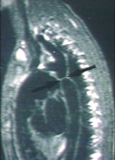
[[Image:Lossless-page1-230px-1532-429X-13-51-2.tiff.png|left|thumb|250px|Aortic coarctation. A. 'Black-blood' oblique sagittal view showing discrete, tight coarctation at the aortic isthmus (arrow). B. 3D, contrast-enhanced CT angiogram showing mildly narrowed bare metal stent (arrow) that partially overlies the left subclavian artery origin. The arrowhead shows a subtle pseudo-aneurysm at the distal end of the stent. C. 3D, contrast-enhanced MR angiogram showing aortic arch hypoplasia and coarctation with a 'jump' by-pass graft posteriorly (arrow). D. 3D, contrast-enhanced MR angiogram showing large pseudo-aneurysm (arrowhead) after previous patch angioplasty repair. The true lumen is shown posteriorly (arrow).]] | |||
==References== | ==References== | ||
Revision as of 22:09, 10 April 2012

|
Aortic coarctation Microchapters |
|
Diagnosis |
|---|
|
Treatment |
|
Case Studies |
|
Aortic coarctation MRI On the Web |
|
American Roentgen Ray Society Images of Aortic coarctation MRI |
|
Risk calculators and risk factors for Aortic coarctation MRI |
Editor-In-Chief: C. Michael Gibson, M.S., M.D. [1]; Associate Editor-in-Chief: Cafer Zorkun, M.D., Ph.D. [2]
Overview
Magnetic resonance imaging (MRI) can define the location and severity of a coarctation. MRI can also detect associated cardiac abnormalities and is used for serial follow-up after surgical repair or balloon angioplasty. MRI is recommended to look for aneurysm formation following repair of a coarctation. MR angiography has almost completely replaced invasive catheter based techniques for evaluating re coarctation. In adults with untreated coarctation blood often reaches the lower body through collaterals, eg. internal thoracic arteries via. the subclavian arteries. Those can be seen on MR or angiography.
MRI

-
![[Courtesy of RadsWiki and copylefted]](/images/7/72/Coarctation-of-the-aorta-MRI-003.jpg)
[Courtesy of RadsWiki and copylefted]
-
![[Courtesy of RadsWiki and copylefted]](/images/c/c6/Coarctation-of-the-aorta-MRI-002.jpg)
[Courtesy of RadsWiki and copylefted]
-
![[Courtesy of RadsWiki and copylefted]](/index.php/File:Coarctation-of-the-aorta-MRI-003.jpg)
![[Courtesy of RadsWiki and copylefted]](/index.php/File:Coarctation-of-the-aorta-MRI-002.jpg)

