Aneurysmal bone cyst: Difference between revisions
m (Bot: Automated text replacement (-{{SIB}} + & -{{EH}} + & -{{EJ}} + & -{{Editor Help}} + & -{{Editor Join}} +)) |
No edit summary |
||
| Line 1: | Line 1: | ||
__NOTOC__ | __NOTOC__ | ||
{{CMG}} | {{CMG}}; {{AE}} {{Rohan}}; {{CZ}} | ||
Revision as of 16:57, 20 December 2018
Editor-In-Chief: C. Michael Gibson, M.S., M.D. [1]; Associate Editor(s)-in-Chief: Rohan A. Bhimani, M.B.B.S., D.N.B., M.Ch.[2]; Cafer Zorkun, M.D., Ph.D. [3]
Overview
An aneurysmal bone cyst is an expansile osteolytic lesion with a thin wall, containing blood-filled cystic cavities. The term aneurysmal is derived from its radiographic appearance.
Causes
Trauma is considered the initiating factor in the development of some cysts, in documented cases involving an acute fracture. Local alterations in the blood flow are related to obstructions that are important in the development of an aneurysmal bone cyst.
The lesion generally arises in a pre-existing bone tumor, this is because of the abnormal bones changes in hemodynamics. An aneurysmal bone cyst can arise from a pre-existing chondroblastoma, a chondromyxoid fibroma, an osteoblastoma, a giant cell tumor, or fibrous dysplasia. A giant cell tumor is the most common cause, occurring in 19% to 39% of cases. Less frequently, it results from some malignant tumors, such as osteosarcoma, chondrosarcoma, and hemangioendothelioma.
Effects
Aneurysmal bone cysts may be intraosseous, staying inside of the bone marrow. Or they may be extraosseous, developing on the surface of the bone, and extending into the marrow.
Phases
- Initial phase of osteolysis.
- Active growth phase, characterized by destruction of bone tissue.
- Stable stage, where a formation of a bony shell and internal bony septa produce its soap bubble appearance.
- Healing phase where the cyst eventually heals into an irregular dense bony mass.
Occurrences
Aneurysmal bone cysts are more common in females than males. Most of these occur between the ages of 10-30, with about 75% of incidences happening to patients under the age of 20.
Anatomy
The tumor can have an occurrence anywhere that there is bone. Approximate percentages by sites are as shown:
- Skull and mandible (4%)
- Spine (16%)
- Clavicle and ribs (5%)
- Upper extremity (21%)
- Pelvis and sacrum (12%)
- Femur (13%)
- Lower leg (24%)
- Foot (3%)
Diagnosis
The afflicted may have relatively small amounts of pain that will quickly increase in severity over a time period of 6-12 weeks. The skin temperature around the bone may increase, a bony swelling may be evident, and movement may be restricted in adjacent joints.
Spinal lesions may cause quadriplegia and patients with skull lesions may have headaches.
The imaging findings are
- Aneurysmal bone cysts are well-defined, cortically based, rapidly expansile lytic lesions.
- The location is usually metaphyseal, and the epicenter is eccentric.
- Long tubular bones and the spine account for 60%–70% of cases.
- They can grow large enough to involve the medullary cavity, although their tendency is for eccentric expansion (a so-called blister lesion).
- Extremely rapid growth is characteristic of aneurysmal bone cysts, as expansion has more to do with vascular engorgement than with cellular proliferation, and aids in its differentiation from other eccentric, expansile lesions of bone.
- Must be differentiated from telangiectatic osteosarcoma.
Patient #1: Radiograph demonstrates an aneurysmal bone cyst in the hand
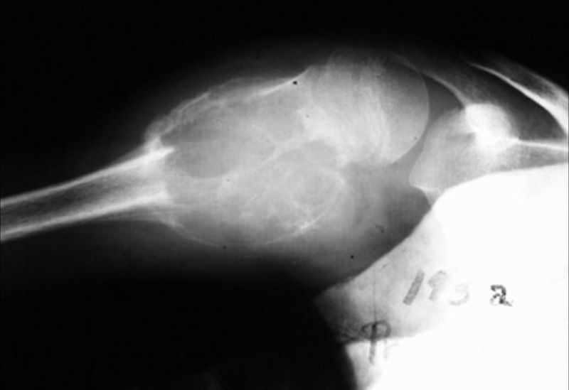
Patient #2: Radiograph demonstrates an aneurysmal bone cyst in the proximal humerus
See also
External links
Template:Diseases of the musculoskeletal system and connective tissue

