Amoebiasis causes
|
Amoebiasis Microchapters |
|
Diagnosis |
|---|
|
Treatment |
|
Case Studies |
|
Amoebiasis causes On the Web |
|
American Roentgen Ray Society Images of Amoebiasis causes |
Editor-In-Chief: C. Michael Gibson, M.S., M.D. [1]
Overview
Entamoeba histolytica can live in the large intestine (colon) without causing disease. However, sometimes, it invades the colon wall, causing colitis, acute dysentery, or long-term (chronic) diarrhea. The infection can also spread through the blood to the liver and, rarely, to the lungs, brain or other organs.
Amoebiasis can be seen anywhere in the world, but it is most common in tropical areas with crowded living conditions and poor sanitation. Africa, Mexico, parts of South America, and India have significant health problems associated with this disease. Entamoeba histolytica is spread through food or water contaminated with stools. This is common when human waste is used as fertilizer. It can also be spread from person to person -- particularly by contact with the mouth or rectal area of an infected person.
Life Cycle
Cysts and trophozoites are passed in feces (1). Cysts are typically found in formed stool, whereas trophozoites are typically found in diarrheal stool. Infection by Entamoeba histolytica occurs by ingestion of mature cysts (2) in fecally contaminated food, water, or hands. Excystation (3) occurs in the small intestine and trophozoites (4) are released, which migrate to the large intestine. The trophozoites multiply by binary fission and produce cysts (5), and both stages are passed in the feces (1). Because of the protection conferred by their walls, the cysts can survive days to weeks in the external environment and are responsible for transmission. Trophozoites passed in the stool are rapidly destroyed once outside the body, and if ingested would not survive exposure to the gastric environment. In many cases, the trophozoites remain confined to the intestinal lumen (A: noninvasive infection) of individuals who are asymptomatic carriers, passing cysts in their stool. In some patients the trophozoites invade the intestinal mucosa (B: intestinal disease), or, through the bloodstream, extraintestinal sites such as the liver, brain, and lungs (C: extraintestinal disease), with resultant pathologic manifestations. It has been established that the invasive and noninvasive forms represent two separate species, respectively E. histolytica and E. dispar. These two species are morphologically indistinguishable unless E. histolytica is observed with ingested red blood cells (erythrophagocystosis). Transmission can also occur through exposure to fecal matter during sexual contact (in which case not only cysts, but also trophozoites could prove infective).
-
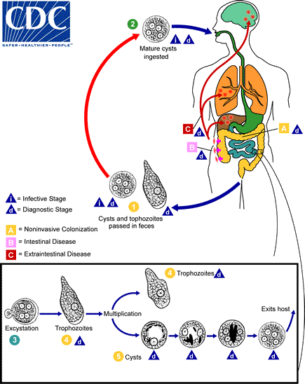
Life cycle of Amebiasis
Adapted from CDC

References