Ventricular action potential
Editor-In-Chief: C. Michael Gibson, M.S., M.D. [1]
Overview
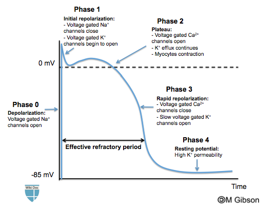
The ventricular action potential is composed of four phases: phase 0 is depolarization, phase 1 is early repolarization, phase 2 is a plateau, phase 3 is rapid repolarization and phase 4 is the resting potential. The different phases depend on the opening and/or closure of specific ion channels.
Ventricular Action Potential
At rest, the ventricular myocyte membrane potential is about -90 mV, which is close to the potassium reversal potential. When an action potential is generated, the membrane potential rises above this level in four distinct phases.
The beginning of the action potential, phase 1, specialized membrane proteins (voltage-gated sodium channels) in the cell membrane selectively allow sodium ions to enter the cell. This causes the membrane potential to rise at a rate of about 300 V/s. As the membrane voltage rises (to about 40 mV) sodium channels close due to a process called inactivation.
The sodium channel opening is followed by inactivation. Sodium inactivation comes with slowly inactivating Ca2+ channels at the same time as a few fast K+ channels open. There is a balance between the centrifugal flow of K+ and the centripetal flow of Ca2+ causing a plateau of length in variables. The delayed opening of more Ca2+-activated K+ channels, which are activated by build-up of Ca2+ in the sarcoplasm, while the Ca2+ channels close, ends the plateau. This leads to repolarisation.
The depolarization of the membrane allows calcium channels to open as well. As sodium channels close calcium provides current to maintain the potential around 10 mV. The plateau lasts on the order of 100 ms. At the time that calcium channels are getting activated, channels that mediate the transient outward potassium current open as well. This outward potassium current causes a small dip in membrane potential shortly after depolarization. This current is observed in human and dog action potentials, but not in guinea pig action potentials.
Repolarization is accomplished by channels that open slowly and are mostly activated at the end of the action potential (slow delayed-rectifier channels), and channels that open quickly but are inactivated until the end of the action potential (rapid delayed rectifier channels). Fast delayed rectifier channels open quickly but are shut by inactivation at high membrane potentials. As the membrane voltage begins to drop the channels recover from inactivation and carry current.
Shown below is an image summarizing the phases of the ventricular action potential.
