Cerebral aneurysm
For patient information, click here
| Cerebral aneurysm | |
 | |
|---|---|
| Brain: Berry Aneurysm: Gross, natural color, close-up, an excellent view of typical berry aneurysm located on anterior cerebral artery Image courtesy of Professor Peter Anderson DVM PhD and published with permission © PEIR, University of Alabama at Birmingham, Department of Pathology |
|
Cerebral aneurysm Microchapters |
|
Diagnosis |
|---|
|
Treatment |
|
Case Studies |
|
Cerebral aneurysm On the Web |
|
American Roentgen Ray Society Images of Cerebral aneurysm |
Editor-In-Chief: C. Michael Gibson, M.S., M.D. [1]; Associate Editor(s)-in-Chief: Cafer Zorkun, M.D., Ph.D. [2]; Kalsang Dolma, M.B.B.S.[3]
Synonyms and keywords: Berry aneurysm
MRI
Images shown below are courtesy of RadsWiki and copylefted.
-
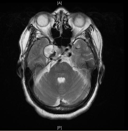
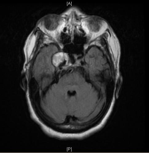
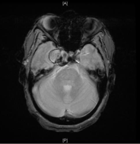
MRI: A large cavernous sinus aneurysm
-
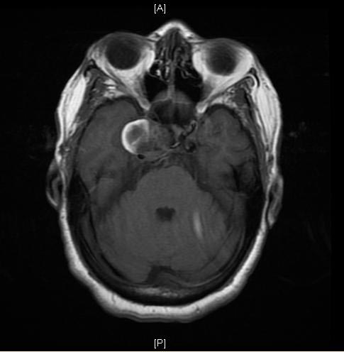
MRI: A large cavernous sinus aneurysm
-
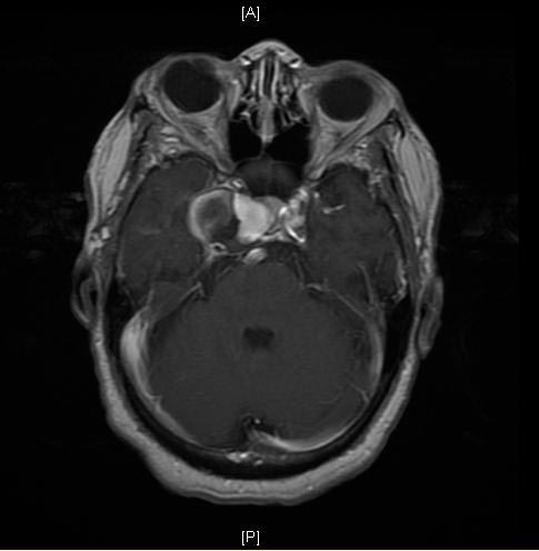
MRI: A large cavernous sinus aneurysm
Angiography
Images shown below are courtesy of RadsWiki and copylefted.
-
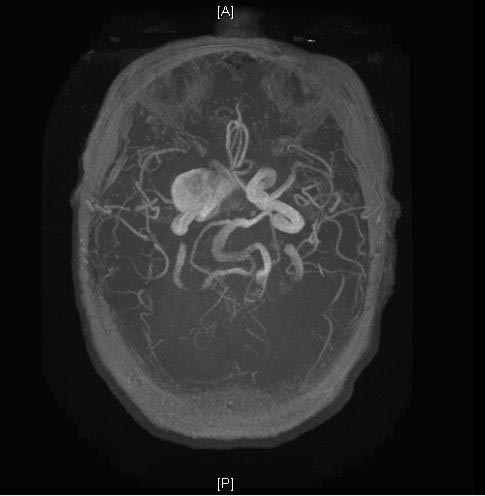
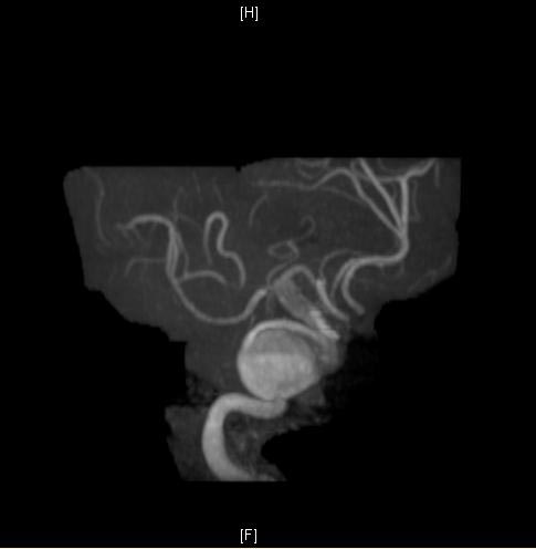
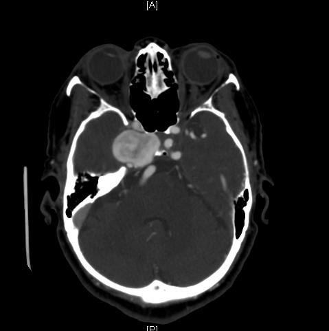
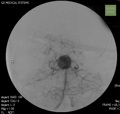
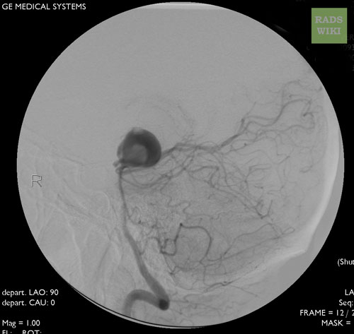
Cranial Angiography: Same case as in MSCT images. A large basilar artery aneurysm
-
Cranial Angiography: Same case as in MSCT images. A large basilar artery aneurysm
References
See also
- Charcot-Bouchard aneurysm
- Intracranial berry aneurysm
- Stroke
- International Subarachnoid Aneurysm Trial
ca:Aneurisma cerebral de:Aneurysma#Hirn-Aneurysmata fi:Aivoaneurysma
