Supraventricular tachycardia electrocardiogram
|
Supraventricular tachycardia Microchapters |
|
Differentiating Among the Different Types of Supraventricular Tachycardia |
|---|
|
Differentiating Supraventricular Tachycardia from Ventricular Tachycardia |
|
Diagnosis |
|
Treatment |
|
2015 ACC/AHA Guideline Recommendations |
|
Case Studies |
|
Supraventricular tachycardia electrocardiogram On the Web |
|
American Roentgen Ray Society Images of Supraventricular tachycardia electrocardiogram |
|
Directions to Hospitals Treating Supraventricular tachycardia |
|
Risk calculators and risk factors for Supraventricular tachycardia electrocardiogram |
Please help WikiDoc by adding content more here. It's easy! Click here to learn about editing.
Editor-In-Chief: C. Michael Gibson, M.S., M.D. [1]
Overview
Electrocardiogram
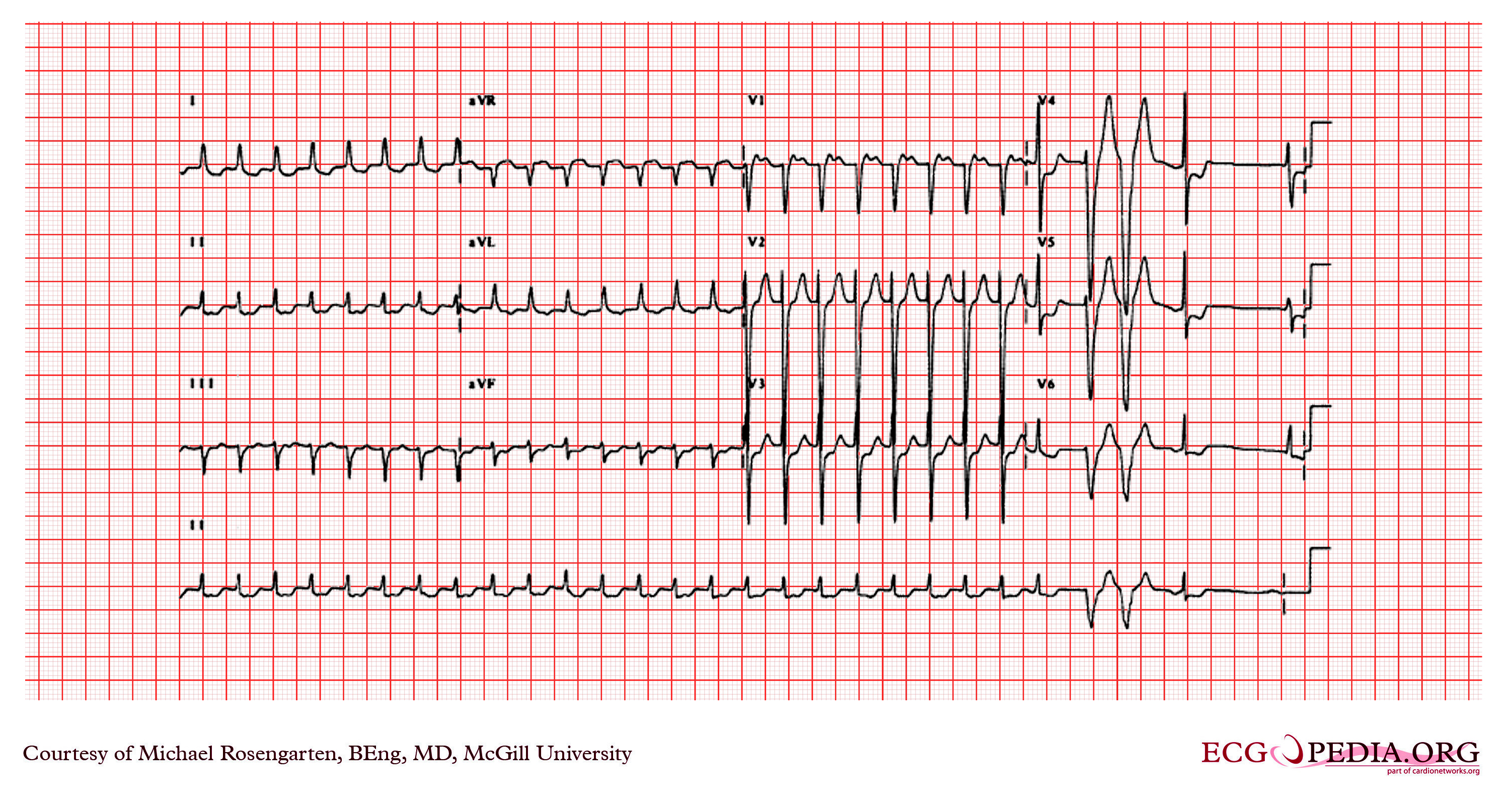
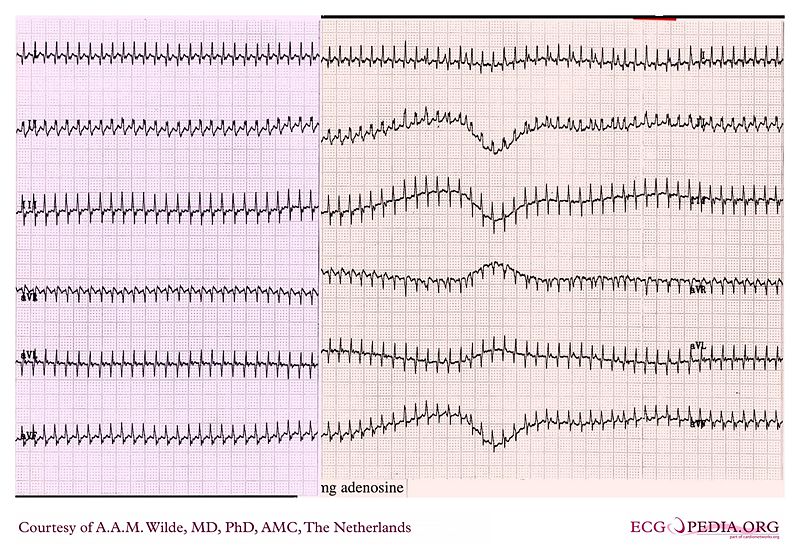
The EKG below is an interesting recording that shows a supraventricular tachycardia. The heart rate is around 185 bpm. It is somewhat unusual presentation for someone with angina. The arrhythmia terminated with adenosine which has a powerful cholinergic effect that blocks conduction through the AV node.

Copyleft image obtained courtesy of ECGpedia, http://en.ecgpedia.org/wiki/File:E345.jpg
The EKG below is an example showing tachycardia at a rate of 190/min with narrow QRS complexes indicating supraventricular tachycardia.

Copyleft image obtained courtesy of ECGpedia, http://en.ecgpedia.org/wiki/File:De-AW00011.jpg
The EKG below is the recording of the patient who goes from sinus rhythm to a wide complex tachycardia at about 130/min. The wide QRS though disappears after nine complexes and is replaced by narrow complexes at a slightly slower rate. No p wave activity is seen. This is a supraventricular tachycardia with a form of abberancy. In this case we are probably seeing a rate dependent left bundle branch block or the effect of a left bundle branch block which persists for the nine complexes because of continued block in the left bundle from the depolarizations from the intact right bundle.

Copyleft image obtained courtesy of ECGpedia, http://en.ecgpedia.org/wiki/Main_Page
The EKG below is an example demonstrating a rapid heart rate at the rate of nearly 300 beats per minute indicating a paroxysmal supraventricular tachycardia.

Copyleft image obtained courtesy of ECGpedia, http://en.ecgpedia.org/wiki/File:De-AW00012.jpg
Shown below is an example of an EKG showing a supraventricular tachycardia with group ventricular beating with clusters of regular rhythm at about 215/min. The regularity and group beating suggest that this is an organized rhythm and not atrial fibrillation. Look carefully at the interval between the 6th and 7th beats in lead II. Clearly atrial activity is seen at about 215/min. This is an interesting case where the diltiazem has slowed down the SVT which has allowed faster conduction down the A/V node and hence an increase in the ventricular rate.

Shown below is the recording shows the intiation of supraventricular tachycardia. There appears to be a p wave on the last part of the last sinus t wave suggesting that this may be an ectopic atrial tachycardia or possibly an atypical form of A/V nodal reentry where one sees the retrograde p wave before the QRS.

Sources
Copyleft images obtained courtesy of ECGpedia, http://en.ecgpedia.org/index.php?title=Special:NewFiles&offset=&limit=500