Cavernous angioma MRI
|
Cavernous angioma Microchapters |
|
Diagnosis |
|---|
|
Treatment |
|
Case Studies |
|
Cavernous angioma MRI On the Web |
|
American Roentgen Ray Society Images of Cavernous angioma MRI |
Editor-In-Chief: C. Michael Gibson, M.S., M.D. [1]<nowiki>; Associate Editor(s)-in-Chief: Edzel Lorraine Co, D.M.D., M.D.
Overview
Diagnosis is most commonly made accidentally by routine magnetic resonance imaging (MRI) screening, but not all MRI exams are created equal. It is paramount that the patient requests a gradient-echo sequence in order to unmask small or punctate lesions which may otherwise remain undetected. These lesions are also more conspicuous on FLAIR imaging compared to standard T2 weighing. FLAIR imaging is different from Gradient sequences, rather, it is similar to T2 weighing but suppresses free-flowing fluid signal. Sometimes quiescent CCMs can be revealed as incidental findings during MRI exams ordered for other reasons.
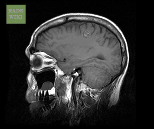
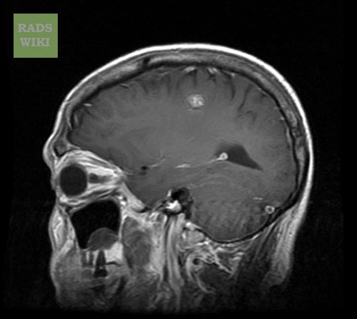
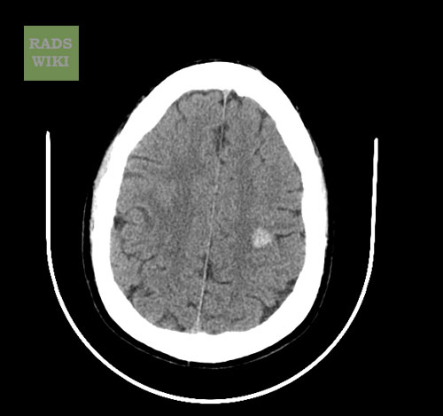
MRI
- Popcorn-like, smoothly circumscribed, well-delineated complex lesions.[1]
- The core is formed by multiple foci of mixed-signal intensities, which represents hemorrhage in various stages of evolution.
- The heterogeneous core typically is surrounded completely by a low-signal-intensity hemosiderin rim on T1-weighted images. The hypointensity of this rim becomes more prominent, or blooms, on T2-weighted and gradient-refocused images because of the magnetic susceptibility effects.
- Smaller cavernous malformations may appear as focal hypointense nodules with both T1- and T2-weighted sequences. The small lesions are depicted more clearly and are more numerous on gradient-echo images because of the increased susceptibility effects of the sequences.
-
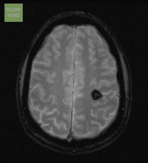
MRI: Cavernous malformation
-
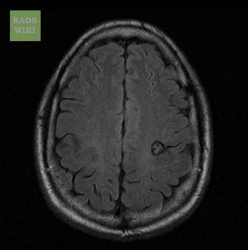
MRI: Cavernous malformation


-
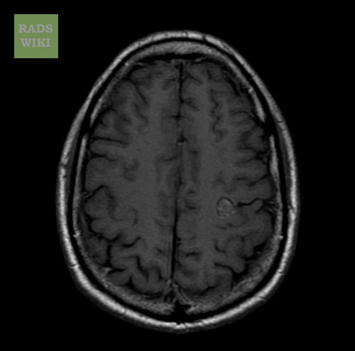
MRI: Cavernous malformation
-
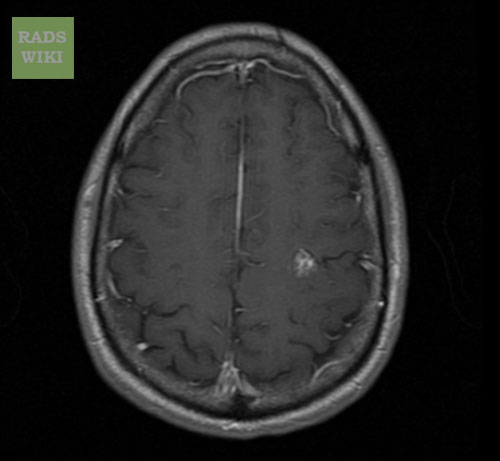
MRI: Cavernous malformation


-
MRI: Cavernous malformation
-
MRI: Cavernous malformation
-
MRI: Cavernous malformation


References
- ↑ Rafee S, Killeen RP, Tubridy N (2021). "'Popcorn' in the Brain: A Cause for Confusion". Am J Med. 134 (2): 216–217. doi:10.1016/j.amjmed.2020.09.014. PMID 33091393 Check
|pmid=value (help).