Seminoma ultrasound
|
Seminoma Microchapters |
|
Diagnosis |
|---|
|
Treatment |
|
Case Studies |
|
Seminoma ultrasound On the Web |
|
American Roentgen Ray Society Images of Seminoma ultrasound |
Editor-In-Chief: C. Michael Gibson, M.S., M.D. [1]Associate Editor(s)-in-Chief: Sujit Routray, M.D. [2]
Overview
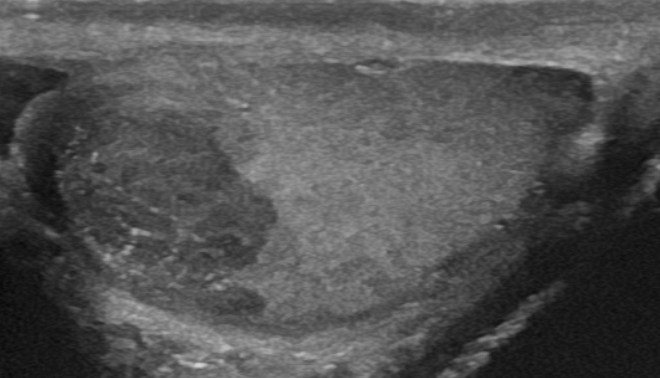
[Imaging study] is the first line imaging modality for seminoma. Findings of seminoma on ultraosund include a homogeneous intratesticular mass of low echogenicity compared to normal testicular tissue.
Ultrasound
- Ultrasound may be helpful in the diagnosis of seminoma. Findings on an ultrasound diagnostic of seminoma include:[1]
- A homogeneous intratesticular mass of low echogenicity compared to normal testicular tissue
- Oval and well-defined mass in the absence of local invasion
- Usually restricted within the tunica albuginea, barely spreading to paratesticular structures
- In color doppler imaging observed internal blood flow
- Heterogeneous appearance can be observed in larger seminoma

References
- ↑ Radiographic features of testicular seminoma. Dr Marcin Czarniecki and Dr Andrew Dixon et al. Radiopaedia 2016. http://radiopaedia.org/articles/testicular-seminoma-1. Accessed on February 25, 2016