Ulnar bone fracture classification
|
Ulnar bone fracture Microchapters |
|
Diagnosis |
|---|
|
Treatment |
|
Case Studies |
|
Ulnar bone fracture classification On the Web |
|
American Roentgen Ray Society Images of Ulnar bone fracture classification |
|
Risk calculators and risk factors for Ulnar bone fracture classification |
Editor-In-Chief: C. Michael Gibson, M.S., M.D. [1]; Associate Editor(s)-in-Chief: Mohammadmain Rezazadehsaatlou[2] ;
Overview
Classification of fractures is an important factor in patients management. Meanwhile the classification of ulnar fracture is important factor in orthopedic medicine.
Classification
- Descriptive[1][2][3][4][5][6]
- closed versus open
- location
- comminuted, segmental, multifragmented
- displacement
- angulation
- rotational alignment
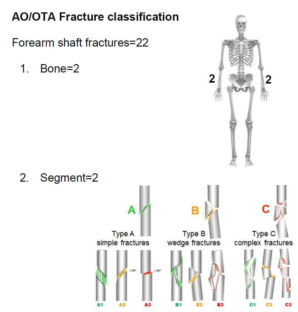
- OTA classification
- radial and ulna diaphyseal fractures
- Type A
- simple fracture of ulna (A1), radius (A2), or both bones (A3)
- Type B
- wedge fracture of ulna (B1), radius (B2), or both bones (B3)
- Type C
- complex fractures
- Type A
- radial and ulna diaphyseal fractures

Refrences
- ↑ Meena S, Sharma P, Sambharia AK, Dawar A (2014). "Fractures of distal radius: an overview". J Family Med Prim Care. 3 (4): 325–32. doi:10.4103/2249-4863.148101. PMC 4311337. PMID 25657938.
- ↑ Youlden DJ, Sundaraj K, Smithers C (October 2018). "Volar locking plating versus percutaneous Kirschner wires for distal radius fractures in an adult population: a meta-analysis". ANZ J Surg. doi:10.1111/ans.14903. PMID 30354018.
- ↑ Atanelov Z, Bentley TP. PMID 30020651. Missing or empty
|title=(help) - ↑ Kiel J, Kaiser K. PMID 29939612. Missing or empty
|title=(help) - ↑ Carter KR, Nallamothu SV. PMID 29763211. Missing or empty
|title=(help) - ↑ Attum B, Thompson JH. PMID 29489190. Missing or empty
|title=(help)