Gout x ray
|
Gout Microchapters |
|
Diagnosis |
|---|
|
Treatment |
|
Case Studies |
|
Gout x ray On the Web |
|
American Roentgen Ray Society Images of Gout x ray |
Editor-In-Chief: C. Michael Gibson, M.S., M.D. [1]
Please help WikiDoc by adding more content here. It's easy! Click here to learn about editing.
Overview
An x-ray is done when gout is suspected to rule out other abnormalities of the bone that may be causing the pain. Most commonly in gout, the x-ray will show no abnormalities, or a small amount of soft tissue swelling.
X-ray
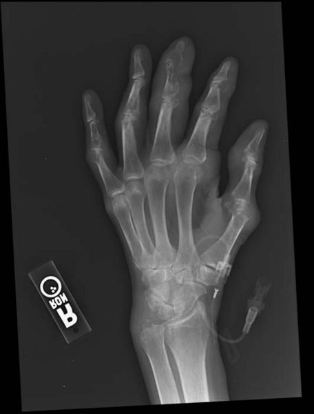
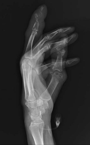
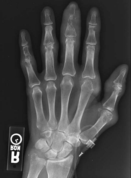
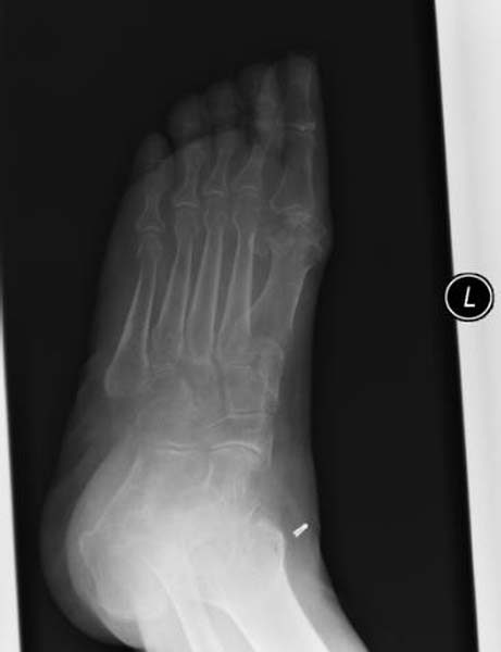
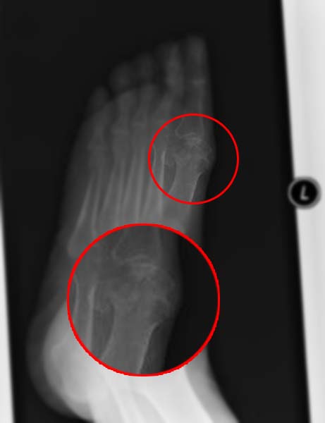
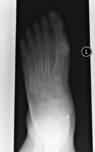
Plain film radiography may be used to evaluate gout; however, radiographic imaging findings generally do not appear until after at least 1 year of uncontrolled disease. The classic radiographic finding of gout late in disease is that of punched-out or rat-bite erosions with overhanging edges and sclerotic margins. null 11
Nuclear medicine studies can be used as a tool to measure the extent of gouty arthritis and to confirm clinically suspected disease. Characteristic findings include increased activity in the affected areas in all phases of a triple-phase bone scan.
Ultrasonography for the diagnosis of gout has advantages in that it is easily available and portable and doesn't require ionizing radiation. However, its limitation include being unable to image deep structures or joint and is highly operator dependent. null 11 Findings include the double-contour sign (hyperechoic irregular enhancement over the surface of the hyaline cartilage) and can identify tophus deposition in and around joints, erosions, and tissue inflammation if power Doppler US is used. [[null 14], [null 15], [null 3], [null 13]] According to the Agency for Healthcare Research and Quality (AHRQ), 4 ultrasound studies on gout showed sensitivities that ranged from 37-100% and specificities that ranged from 68-97%. null 12
CT scanning can be used to study the effects of gout in areas that are hard to visualize with plain-film radiography. Many studies have been performed using dual-energy CT with good results, providing visualization, characterization, and quantification of monosodium urate crystals. [[null 16], [null 17], [null 18], [null 11], [null 19], [null 4]] DECT scanners are able to perform simultaneous acquisitions at 80 and 140 kVp using two separate sets of x‐ray tubes and detectors positioned 90 to 95 degrees apart, thereby differentiating materials based on their relative absorption of x‐rays at the different photon energy levels. null 11 According to the AHRQ, DECT has shown good sensitivity and specificity for predicting gout compared with synovial fluid analysis for monosodium urate crystals, with 3 studies showing sensitivities that ranged from 85-100% and specificities that ranged from 83-92%.
The goal of joint X Ray is to rule out other diseases that affect the joint. The most common radiographic findings in patients with gout include soft-tissue swelling or an absence of abnormalities.
Patient #1
Patient #2
Sources
Copyleft images obtained courtesy of RadsWiki [2]
