Giardia lamblia
|
Giardiasis Microchapters |
|
Diagnosis |
|---|
|
Treatment |
|
Case Studies |
|
Giardia lamblia On the Web |
|
American Roentgen Ray Society Images of Giardia lamblia |
| Giardia lamblia | ||||||||
|---|---|---|---|---|---|---|---|---|
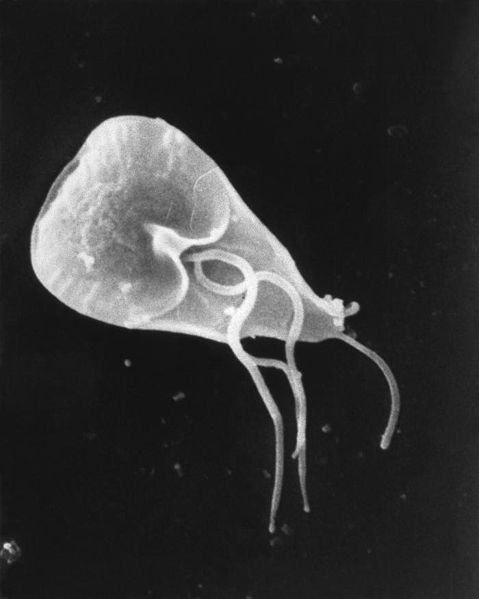
 Giardia cell, SEM
| ||||||||
| Scientific classification | ||||||||
| ||||||||
| Binomial name | ||||||||
| Giardia lamblia (Kunstler, 1882) |
This page is about microbiologic aspects of the organism(s). For clinical aspects of the disease, see Giardiasis.
Editor-In-Chief: C. Michael Gibson, M.S., M.D. [1]
Overview
Giardia lamblia (synonymous with Lamblia intestinalis and Giardia duodenalis) is a flagellated protozoan parasite that is responsible for the development of giardiasis.
Higher Order Classification
Eukaryota, Diplomonadida group, Diplomonadida, Hexamitidae, Giardiinae, Giardia, G. lamblia
Natural Reservoir
- Giardia affects humans and animals, such as cats, dogs, cows, beavers, deer, and sheep.
Microbiological Characteristicsc
- Giardia lamblia is a flagellated, microaerophilic parasite.
- The trophozoite form of G. lamblia is pear-shaped and has a unique morphology that includes two identical nuclei, a ventral disc for adhesion to the host intestine, and flagella.
Genome
- G. lamblia genome consists of 1.2 million base pairs (average GC content: 46%).[1]
- The genome pairs are distributed across five linear chromosomes.[1]
- Similar to other eukaryotes, each chromosome is flanked by the telomere sequence (5’TAGGG3’).[1]
Life cycle

Giardia belongs among the diplomonads.
Non-infective Cyst
- The life cycle begins with a noninfective cyst being excreted with faeces of an infected individual. Once out in the environment, the cyst becomes infective.
- A distinguishing characteristic of the cyst is 4 nuclei and a retracted cytoplasm.
Trophozoite
- Once ingested by a host, the trophozoite emerges to an active state of feeding and motility.
- After the feeding stage, the trophozoite undergoes asexual replication through longitudinal binary fission.
- The resulting trophozoites and cysts then pass through the digestive system in the feces.
- While the trophozoites may be found in the feces, only the cysts are capable of surviving outside of the host.
- Distinguishing features of the trophozoites are large karyosomes and lack of peripheral chromatin, giving the two nuclei a halo appearance.
-
SEM depicts the dorsal surface of a Giardia protozoan, isolated from a rat’s intestine. From Public Health Image Library (PHIL). [2]
-
SEM depicts the mucosal surface of the small intestine of a gerbil infested with Giardia sp. protozoa. From Public Health Image Library (PHIL). [2]
-
SEM depicts a Giardia lamblia protozoan in a late stage of cell division that was about to become two separate organisms, producing a heart-shaped form. From Public Health Image Library (PHIL). [2]
-
SEM depicts the ventral surface of a Giardia muris trophozoite. From Public Health Image Library (PHIL). [2]
-
SEM depicts dorsal surface of a Giardia protozoan, isolated from a rat’s intestine. From Public Health Image Library (PHIL). [2]
-
SEM depicts some of the ultrastructural morphologic details of an oblong-shaped Giardia sp. protozoan cyst. From Public Health Image Library (PHIL). [2]
-
SEM depicts the ventral surface of a Giardia muris trophozoite that had settled atop the mucosal surface of a rat’s intestine. From Public Health Image Library (PHIL). [2]
-
SEM depicts a Giardia lamblia protozoan that was about to become two separate organisms, as it was caught in a late stage of cell division. From Public Health Image Library (PHIL). [2]
-
SEM depicts a Giardia muris protozoan adhering itself to the microvillous border of an intestinal epithelial cell. From Public Health Image Library (PHIL). [2]
-
This photomicrograph depicts Giardia lamblia parasites using indirect immunofluorescence test for giardiasis. From Public Health Image Library (PHIL). [2]
References
- ↑ 1.0 1.1 1.2 Le Blancq SM, Kase RS, Van der Ploeg LH (1991). "Analysis of a Giardia lamblia rRNA encoding telomere with [TAGGG]n as the telomere repeat". Nucleic Acids Res. 19 (20): 5790. PMC 328996. PMID 1840670.
- ↑ 2.00 2.01 2.02 2.03 2.04 2.05 2.06 2.07 2.08 2.09 "Public Health Image Library (PHIL)".
![SEM depicts the dorsal surface of a Giardia protozoan, isolated from a rat’s intestine. From Public Health Image Library (PHIL). [2]](/images/b/b7/Giardiasis09.jpeg)
![SEM depicts the mucosal surface of the small intestine of a gerbil infested with Giardia sp. protozoa. From Public Health Image Library (PHIL). [2]](/images/3/36/Giardiasis08.jpeg)
![SEM depicts a Giardia lamblia protozoan in a late stage of cell division that was about to become two separate organisms, producing a heart-shaped form. From Public Health Image Library (PHIL). [2]](/images/2/24/Giardiasis07.jpeg)
![SEM depicts the ventral surface of a Giardia muris trophozoite. From Public Health Image Library (PHIL). [2]](/images/0/05/Giardiasis06.jpeg)
![SEM depicts dorsal surface of a Giardia protozoan, isolated from a rat’s intestine. From Public Health Image Library (PHIL). [2]](/images/e/e5/Giardiasis05.jpeg)
![SEM depicts some of the ultrastructural morphologic details of an oblong-shaped Giardia sp. protozoan cyst. From Public Health Image Library (PHIL). [2]](/images/1/1c/Giardiasis04.jpeg)
![SEM depicts the ventral surface of a Giardia muris trophozoite that had settled atop the mucosal surface of a rat’s intestine. From Public Health Image Library (PHIL). [2]](/images/9/9e/Giardiasis03.jpeg)
![SEM depicts a Giardia lamblia protozoan that was about to become two separate organisms, as it was caught in a late stage of cell division. From Public Health Image Library (PHIL). [2]](/images/2/2e/Giardiasis02.jpeg)
![SEM depicts a Giardia muris protozoan adhering itself to the microvillous border of an intestinal epithelial cell. From Public Health Image Library (PHIL). [2]](/images/f/f4/Giardiasis01.jpeg)
![This photomicrograph depicts Giardia lamblia parasites using indirect immunofluorescence test for giardiasis. From Public Health Image Library (PHIL). [2]](/images/5/55/Giardiasis10.jpeg)