WBR0042: Difference between revisions
Jump to navigation
Jump to search
Rim Halaby (talk | contribs) No edit summary |
Rim Halaby (talk | contribs) No edit summary |
||
| Line 25: | Line 25: | ||
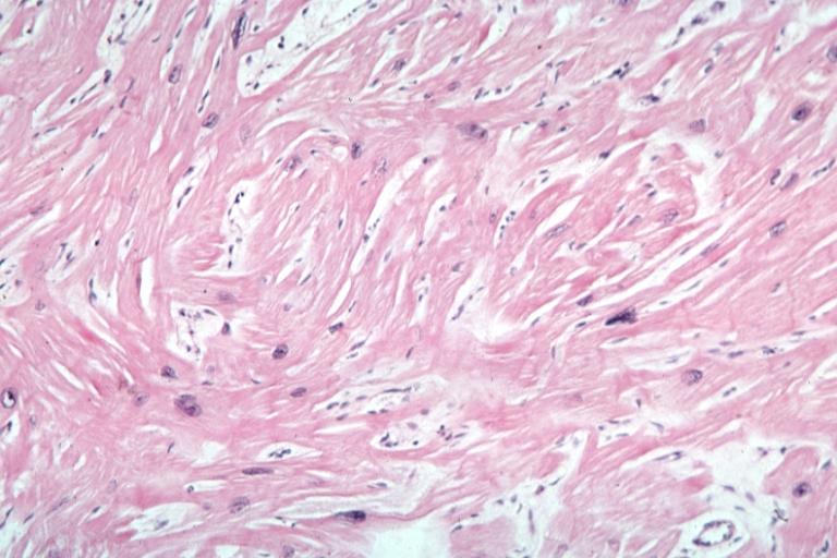
#'''Histologically''' there are hypertrophied [[cardiomyocytes]] in disarray which alters the conduction system and subsequently predisposes to [[arrhythmias]]. [[File:438.jpg|center|200px]] | #'''Histologically''' there are hypertrophied [[cardiomyocytes]] in disarray which alters the conduction system and subsequently predisposes to [[arrhythmias]]. [[File:438.jpg|center|200px]] | ||
|AnswerA=Symmetric left ventricular hypertrophy | |AnswerA=Symmetric left ventricular hypertrophy | ||
|AnswerAExp=Symmetric left ventricular hypertrophy is characteristically present in patients with increased afterload, such as [[ | |AnswerAExp=Symmetric left ventricular hypertrophy is characteristically present in patients with increased [[afterload]], such as [[aortic stenosis]] or [[hypertension]]. This leads to the increased synthesis of [[actin]] and [[myosin]] that are arranged in a "organized fashion". Ultimately the patient develops diastolic dysfunction due to the inability of the heart to fill in with blood during [[diastole]]. | ||
|AnswerB=White appearance of the endocardium | |AnswerB=White appearance of the endocardium | ||
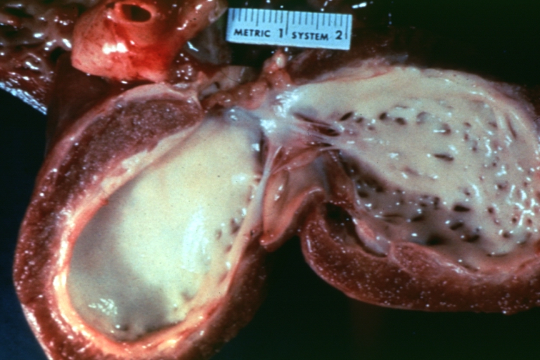
|AnswerBExp= | |AnswerBExp=A white appearance of the [[endocardium]] can be present in [[endocardial fibroelastosis]], a rare restrictive [[cardiomyopathy]] present in young children less than 2 years old. It is due to an excessive [[fibrosis]] of the [[endocardium]] that causes diastolic dysfunction. | ||
[[Image:Endocardial_fibroelastosis_2.jpg|center|200px]] | [[Image:Endocardial_fibroelastosis_2.jpg|center|200px]] | ||
|AnswerC=Cardiomyocytes hypertrophy in an organized fashion | |AnswerC=Cardiomyocytes hypertrophy in an organized fashion | ||
|AnswerCExp= | |AnswerCExp=Cardiomyocytes hypertrophy in an organized fashion is present among patients with increased [[afterload]] such as [[aortic stenosis]] or [[hypertension]]. | ||
|AnswerD=Prominent ventricular septum hypertrophy compared to the ventricular wall | |AnswerD=Prominent ventricular septum hypertrophy compared to the ventricular wall | ||
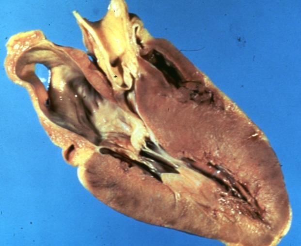
|AnswerE=Fibrotic thickening of endocardium and valves of the right side of the heart | |AnswerDExp=Prominent ventricular septum hypertrophy is a characteristic finding of [[hypertrophic cardiomyopathy]]. | ||
|AnswerEExp= | |AnswerE=Fibrotic thickening of the endocardium and the valves of the right side of the heart | ||
|AnswerEExp=[[Fibrosis|Fibrotic thickening]] of the [[endocardium]] and the valves of the right side of the heart is the macroscopic description of an endocardium affected by a [[carcinoid syndrome|carcinoid syndrome]] due to chronic [[serotonin]] exposure, which causes fibrosis of the tricuspid valve and pulmonary valve. [[Carcinoid syndrome]] occurs when the [[carcinoid tumor]] metastasize to the liver. The patient clinically presents with [[diarrhea]], [[wheezing]], [[telangiectasias]], and [[flushing]] of the skin. | |||
|EducationalObjectives=Hypertrophic obstructive cardiomyopathy (HOCM) is a common cause of sudden death in young athletes during intense exercise. It is characterized by the presence of cardiac hypertrophy more prominent in the ventricular septum and hypertrophied [[cardiomyocytes]] in disarray. | |EducationalObjectives=Hypertrophic obstructive cardiomyopathy (HOCM) is a common cause of sudden death in young athletes during intense exercise. It is characterized by the presence of cardiac hypertrophy more prominent in the ventricular septum and hypertrophied [[cardiomyocytes]] in disarray. | ||
|RightAnswer=D | |RightAnswer=D | ||
Revision as of 19:32, 15 March 2014
| Author | PageAuthor::Gonzalo Romero |
|---|---|
| Exam Type | ExamType::USMLE Step 1 |
| Main Category | MainCategory::Pathology |
| Sub Category | SubCategory::Cardiology |
| Prompt | [[Prompt::A 21-year-old healthy male college student is playing in the football finale game across local colleges when he suddenly falls on the ground while running. The player is found unresponsive. The Emergency Medical Services arrive promptly and initiate CPR and resuscitation measures without success. According to his family and friends, he has always been healthy and playing football since high school. Autopsies are obtained in order to determine the cause of death. Which of the following cardiac macroscopic or microscopic changes is most likely to be present?]] |
| Answer A | AnswerA::Symmetric left ventricular hypertrophy |
| Answer A Explanation | [[AnswerAExp::Symmetric left ventricular hypertrophy is characteristically present in patients with increased afterload, such as aortic stenosis or hypertension. This leads to the increased synthesis of actin and myosin that are arranged in a "organized fashion". Ultimately the patient develops diastolic dysfunction due to the inability of the heart to fill in with blood during diastole.]] |
| Answer B | AnswerB::White appearance of the endocardium |
| Answer B Explanation | [[AnswerBExp::A white appearance of the endocardium can be present in endocardial fibroelastosis, a rare restrictive cardiomyopathy present in young children less than 2 years old. It is due to an excessive fibrosis of the endocardium that causes diastolic dysfunction.
 |
| Answer C | AnswerC::Cardiomyocytes hypertrophy in an organized fashion |
| Answer C Explanation | [[AnswerCExp::Cardiomyocytes hypertrophy in an organized fashion is present among patients with increased afterload such as aortic stenosis or hypertension.]] |
| Answer D | AnswerD::Prominent ventricular septum hypertrophy compared to the ventricular wall |
| Answer D Explanation | [[AnswerDExp::Prominent ventricular septum hypertrophy is a characteristic finding of hypertrophic cardiomyopathy.]] |
| Answer E | AnswerE::Fibrotic thickening of the endocardium and the valves of the right side of the heart |
| Answer E Explanation | [[AnswerEExp::Fibrotic thickening of the endocardium and the valves of the right side of the heart is the macroscopic description of an endocardium affected by a carcinoid syndrome due to chronic serotonin exposure, which causes fibrosis of the tricuspid valve and pulmonary valve. Carcinoid syndrome occurs when the carcinoid tumor metastasize to the liver. The patient clinically presents with diarrhea, wheezing, telangiectasias, and flushing of the skin.]] |
| Right Answer | RightAnswer::D |
| Explanation | [[Explanation::This young athlete presents with a sudden death during intense exercise due to ventricular arrhythmias, a typical clinical presentation of hypertrophic cardiomyopathy, also known as hypertrophic obstructive cardiomyopathy (HOCM), asymmetrical septal hypertrophy or idiopathic hypertrophic subaortic stenosis (IHSS). Hypertrophic cardiomyopathy can be either autosomal dominant or idiopathic.
Educational Objective: Hypertrophic obstructive cardiomyopathy (HOCM) is a common cause of sudden death in young athletes during intense exercise. It is characterized by the presence of cardiac hypertrophy more prominent in the ventricular septum and hypertrophied cardiomyocytes in disarray. |
| Approved | Approved::Yes |
| Keyword | WBRKeyword::Cardiology, WBRKeyword::Pathology, WBRKeyword::Hypertrophic cardiomyopathy, WBRKeyword::HOCM |
| Linked Question | Linked:: |
| Order in Linked Questions | LinkedOrder:: |
Image [[WBRImage::|]] Caption WBRImageCaption::no-display Position [[WBRImagePlace::|]]

