Aortic coarctation classification: Difference between revisions
| Line 10: | Line 10: | ||
There are three variations of coarctation of the aorta:<ref>Valdes-Cruz LM, Cayre RO: ''Echocardiographic diagnosis of congenital heart disease.'' Philadelphia, 1998.</ref> | There are three variations of coarctation of the aorta:<ref>Valdes-Cruz LM, Cayre RO: ''Echocardiographic diagnosis of congenital heart disease.'' Philadelphia, 1998.</ref> | ||
==Preductal coarctation== | |||
The narrowing is proximal to the ductus arteriosus. If severe, blood flow to the aorta distal (to lower body) to the narrowing is dependent on a patent ductus arteriosus, and hence its closure can be life-threatening. Preductal coarctation results when an intracardiac anomaly during fetal life decreases blood flow through the left side of the heart, leading to hypoplastic development of the aorta. | |||
==Ductal coarctation== | |||
The narrowing occurs at the insertion of the ductus arteriosus. This kind usually appears when the ductus arteriosus closes. | |||
==Postductal coarctation== | |||
The narrowing is distal to the insertion of the ductus arteriosus. Even with an open ductus arteriosus blood flow to the lower body can be impaired. Newborns with this type of coarctation may be critically sick from the birth. This type is most common in adults. It is associated with notching of the ribs, hypertension in the upper extremities, and weak pulses in the lower extremities. Postductal coarctation is most likely the result of muscular ductal (ductus arteriosis) extends into the aorta during fetal life | |||
==References== | ==References== | ||
Revision as of 01:27, 18 October 2012
|
Aortic coarctation Microchapters |
|
Diagnosis |
|---|
|
Treatment |
|
Case Studies |
|
Aortic coarctation classification On the Web |
|
American Roentgen Ray Society Images of Aortic coarctation classification |
|
Risk calculators and risk factors for Aortic coarctation classification |
Editor-In-Chief: C. Michael Gibson, M.S., M.D. [1]; Associate Editor(s)-In-Chief: Priyamvada Singh, M.B.B.S.[2], Cafer Zorkun, M.D., Ph.D. [3]; Assistant Editor(s)-In-Chief: Kristin Feeney, B.S.[4]
Overview
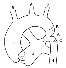
Aortic coarctation can be classified as preductal coarctation, ductal coarctation, and postductal coarctation depending upon the coarctation's anatomic relationship to the ductus arteriosus. All classifications involve narrowings of the aorta that directly impact the aortic hemodynamics.
Classification

There are three variations of coarctation of the aorta:[1]
Preductal coarctation
The narrowing is proximal to the ductus arteriosus. If severe, blood flow to the aorta distal (to lower body) to the narrowing is dependent on a patent ductus arteriosus, and hence its closure can be life-threatening. Preductal coarctation results when an intracardiac anomaly during fetal life decreases blood flow through the left side of the heart, leading to hypoplastic development of the aorta.
Ductal coarctation
The narrowing occurs at the insertion of the ductus arteriosus. This kind usually appears when the ductus arteriosus closes.
Postductal coarctation
The narrowing is distal to the insertion of the ductus arteriosus. Even with an open ductus arteriosus blood flow to the lower body can be impaired. Newborns with this type of coarctation may be critically sick from the birth. This type is most common in adults. It is associated with notching of the ribs, hypertension in the upper extremities, and weak pulses in the lower extremities. Postductal coarctation is most likely the result of muscular ductal (ductus arteriosis) extends into the aorta during fetal life
References
- ↑ Valdes-Cruz LM, Cayre RO: Echocardiographic diagnosis of congenital heart disease. Philadelphia, 1998.