Reticulocyte: Difference between revisions
No edit summary |
Varun Kumar (talk | contribs) |
||
| Line 24: | Line 24: | ||
==Differential Diagnosis of Reticulocytosis== | ==Differential Diagnosis of Reticulocytosis== | ||
{|style="width:90%; height:100px" border="1" | |||
|style="height:100px"; style="width:25%" border="1" bgcolor="LightSteelBlue" | '''Cardiovascular''' | |||
|style="height:100px"; style="width:75%" border="1" bgcolor="Beige" | No underlying causes | |||
|- | |||
|-bgcolor="LightSteelBlue" | |||
| '''Chemical / poisoning''' | |||
|bgcolor="Beige"| No underlying causes | |||
|- | |||
|-bgcolor="LightSteelBlue" | |||
| '''Dermatologic''' | |||
|bgcolor="Beige"| No underlying causes | |||
Acute Myelogenous/Blastic Leukemia AML | |- | ||
Leukemia | |-bgcolor="LightSteelBlue" | ||
Lymphoma/malignant, non-Hodgkins | | '''Drug Side Effect''' | ||
|bgcolor="Beige"| No underlying causes | |||
Primary Myelofibrosis/Myeloid metaplasia | |- | ||
Metabolic | |-bgcolor="LightSteelBlue" | ||
Impaired folic acid metabolism | | '''Ear Nose Throat''' | ||
|bgcolor="Beige"| No underlying causes | |||
Anemia of malnutrition | |- | ||
Folate depletion | |-bgcolor="LightSteelBlue" | ||
Folic acid deficiency anemia | | '''Endocrine''' | ||
Inadequate Folic acid in diet | |bgcolor="Beige"| No underlying causes | ||
Iron deficiency anemia | |- | ||
Iron deficient diet | |-bgcolor="LightSteelBlue" | ||
Malnutrition/Starvation | | '''Environmental''' | ||
Folic acid dependency/metabolic defect | |bgcolor="Beige"| No underlying causes | ||
Kwashiorkor (protein deficiency,severe) | |- | ||
Malabsorption of folic acid | |-bgcolor="LightSteelBlue" | ||
Pellagra/niacin deficiency | | '''Gastroenterologic''' | ||
Vitamin B12 deficiency | |bgcolor="Beige"| No underlying causes | ||
|- | |||
|-bgcolor="LightSteelBlue" | |||
| '''Genetic''' | |||
|bgcolor="Beige"| No underlying causes | |||
|- | |||
|-bgcolor="LightSteelBlue" | |||
| '''Hematologic''' | |||
|bgcolor="Beige"| [[AML|Acute Myelogenous/Blastic Leukemia]] [[AML]], [[Leukemia]], [[Lymphoma]]/malignant, [[Myeloproliferative disease]], [[non-Hodgkins lymphoma]], Primary [[Myelofibrosis]]/[[Myeloid metaplasia]], [[Folate]] depletion, [[Folic acid deficiency]] anemia, [[Iron deficiency anemia]], [[Pellagra]]/[[niacin deficiency]], [[Vitamin B12 deficiency]] | |||
|- | |||
|-bgcolor="LightSteelBlue" | |||
| '''Iatrogenic''' | |||
|bgcolor="Beige"| No underlying causes | |||
|- | |||
|-bgcolor="LightSteelBlue" | |||
Addison's disease (chronic adrenal | | '''Infectious Disease''' | ||
|bgcolor="Beige"| [[Abscesses]], [[Bacteremia]]/[[Septicemia]], Infected organ, [[Parvovirus]] Infection/[[Parvovirus B19]], Posthepatitic [[aplastic anemia]], [[Tuberculosis]] of bone marrow | |||
Adrenocorticoid (Isolated) Deficiency | |- | ||
|-bgcolor="LightSteelBlue" | |||
| '''Musculoskeletal / Ortho''' | |||
Anemia of chronic disease | |bgcolor="Beige"| No underlying causes | ||
Anemia of | |- | ||
Anemia | |-bgcolor="LightSteelBlue" | ||
| '''Neurologic''' | |||
|bgcolor="Beige"| No underlying causes | |||
|- | |||
Aplastic anemia crisis | |-bgcolor="LightSteelBlue" | ||
| '''Nutritional / Metabolic''' | |||
|bgcolor="Beige"| Impaired [[folic acid]] metabolism, [[Anemia of malnutrition]], [[Folate]] depletion, [[Folic acid deficiency]] anemia, Inadequate Folic acid in diet, [[Iron deficiency anemia]], Iron deficient diet, [[Malnutrition]]/Starvation, Folic acid dependency/metabolic defect, [[Kwashiorkor]] (protein deficiency,severe), Malabsorption of folic acid, [[Pellagra]]/[[niacin deficiency]], [[Vitamin B12 deficiency]] | |||
|- | |||
|-bgcolor="LightSteelBlue" | |||
Combined system disease/pernicious | | '''Obstetric/Gynecologic''' | ||
|bgcolor="Beige"| No underlying causes | |||
|- | |||
|-bgcolor="LightSteelBlue" | |||
| '''Oncologic''' | |||
|bgcolor="Beige"| [[AML|Acute Myelogenous/Blastic Leukemia]] [[AML]], [[Leukemia]], [[Lymphoma]]/malignant, [[Myeloproliferative disease]], [[non-Hodgkins lymphoma]], Primary [[Myelofibrosis]]/[[Myeloid metaplasia]] | |||
|- | |||
|-bgcolor="LightSteelBlue" | |||
Methotrexate (Rheumatrex) Administration/Toxicity | | '''Opthalmologic''' | ||
|bgcolor="Beige"| No underlying causes | |||
|- | |||
|-bgcolor="LightSteelBlue" | |||
| '''Overdose / Toxicity''' | |||
|bgcolor="Beige"| No underlying causes | |||
|- | |||
Tetraethyl lead poisoning | |-bgcolor="LightSteelBlue" | ||
| '''Psychiatric''' | |||
|bgcolor="Beige"| No underlying causes | |||
|- | |||
|-bgcolor="LightSteelBlue" | |||
| '''Pulmonary''' | |||
|bgcolor="Beige"| No underlying causes | |||
|- | |||
|-bgcolor="LightSteelBlue" | |||
| '''Renal / Electrolyte''' | |||
|bgcolor="Beige"| No underlying causes | |||
|- | |||
|-bgcolor="LightSteelBlue" | |||
| '''Rheum / Immune / Allergy''' | |||
|bgcolor="Beige"| No underlying causes | |||
|- | |||
|-bgcolor="LightSteelBlue" | |||
| '''Sexual''' | |||
|bgcolor="Beige"| No underlying causes | |||
|- | |||
|-bgcolor="LightSteelBlue" | |||
| '''Trauma''' | |||
|bgcolor="Beige"| No underlying causes | |||
|- | |||
|-bgcolor="LightSteelBlue" | |||
| '''Urologic''' | |||
|bgcolor="Beige"| No underlying causes | |||
|- | |||
|-bgcolor="LightSteelBlue" | |||
| '''Miscellaneous''' | |||
|bgcolor="Beige"| No underlying causes | |||
|- | |||
|} | |||
==Differential Diagnosis of Reticulocytosis== | |||
(In alphabetical order) | |||
* Acquired [[sideroblastic anemia]] | |||
* [[Addison's disease]] ([[chronic adrenal insufficiency]]) | |||
* [[Adrenocortical Insufficiency|Adrenocorticoid (Isolated) Deficiency]] | |||
* [[Acute Myelogenous/Blastic Leukemia|AML]] ([[AML]]) | |||
* [[Androgen insensitivity syndrome]] | |||
* [[Anemia of chronic disease]] | |||
* [[Anemia]] of malnutrition | |||
* Anemia of [[uremia]] | |||
* [[Antimetabolite|Antimetabolite medication Administration/Toxicity]] | |||
* [[Aplastic anemia]] | |||
* [[Aplastic anemia]] crisis | |||
* [[Bacteremia]]/[[Septicemia]] | |||
* [[Chemotherapy]], [[cancer]] (anti-neoplastic) | |||
* Chronic [[alcoholism]] | |||
* [[Chronic liver disease]] | |||
* [[Chronic renal failure]] | |||
* Combined system disease/[[pernicious anemia]] | |||
* Congenital [[aplastic anemia]] | |||
* [[Congenital folate malabsorption]] | |||
* [[Drug induced anemia]] | |||
* Drug induced [[Bone marrow suppression]]. | |||
* Fanconi's pancytopenia-dysmelia syndrome | |||
* [[Folate]] depletion | |||
* [[Folic acid deficiency]] anemia | |||
* Folic acid dependency/metabolic defect | |||
* Hemoglobin H disease | |||
* [[Hereditary elliptocytosis]] | |||
* Hereditary [[sideroblastic anemia]] | |||
* [[Hypogonadism]], | |||
* [[Hypothyroidism]] ([[myxedema]]) | |||
* Impaired folic acid metabolism | |||
* Inadequate Folic acid in diet | |||
* Infected organ, Abscesses | |||
* [[Iron deficiency anemia]] | |||
* Iron deficient diet | |||
* [[Klinefelter's syndrome]] | |||
* [[Kwashiorkor]] (severe protein deficiency) | |||
* [[Lead poisoning]] | |||
* [[Leukemia]] | |||
* Liver disease/Liver disorders | |||
* Malabsorption of folic acid | |||
* [[Malnutrition]]/[[Starvation]] | |||
* [[Megaloblastic anemia]] | |||
* [[Megaloblastic anemia]] of [[pregnancy]] | |||
* [[Methotrexate]] ([[Rheumatrex]]) Administration/Toxicity | |||
* [[Myeloid metaplasia]] pathophysiology | |||
* [[Myelophthisic anemia]] | |||
* [[Myeloproliferative disease]] | |||
* [[Non-Hodgkins lymphoma]]/malignant, | |||
* [[Parvovirus]] Infection/[[Parvovirus B19]] | |||
* [[Pellagra]]/[[niacin deficiency]] | |||
* [[Pernicious anemia]] | |||
* Posthepatitic [[aplastic anemia]] | |||
* Postirradiation effects | |||
* Primary [[Myelofibrosis]]/[[Myeloid metaplasia]] | |||
* Radiation exposure | |||
* Refractory [[megaloblastic anemia]] | |||
* Secondary [[myelofibrosis]] | |||
* Severe, acute [[Radiation sickness]] | |||
* [[Sideroblastic Anemia]] Siderochrestic | |||
* Tetraethyl lead poisoning | |||
* [[Thalassemia major]] | |||
* [[Thalassemia minor]] | |||
* Thalassemia-hemoglobin C disease | |||
* [[Tuberculosis]] of bone marrow | |||
* [[Vitamin B12 deficiency]] | |||
==Diagnosis== | ==Diagnosis== | ||
Revision as of 15:36, 15 April 2012
| Reticulocyte | |
 | |
|---|---|
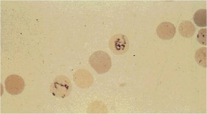
| Peripheral blood; Reticulocyte. © Image courtesy of Nivaldo Medeiros MD and published with permission |
|
WikiDoc Resources for Reticulocyte |
|
Articles |
|---|
|
Most recent articles on Reticulocyte Most cited articles on Reticulocyte |
|
Media |
|
Powerpoint slides on Reticulocyte |
|
Evidence Based Medicine |
|
Clinical Trials |
|
Ongoing Trials on Reticulocyte at Clinical Trials.gov Clinical Trials on Reticulocyte at Google
|
|
Guidelines / Policies / Govt |
|
US National Guidelines Clearinghouse on Reticulocyte
|
|
Books |
|
News |
|
Commentary |
|
Definitions |
|
Patient Resources / Community |
|
Patient resources on Reticulocyte Discussion groups on Reticulocyte Patient Handouts on Reticulocyte Directions to Hospitals Treating Reticulocyte Risk calculators and risk factors for Reticulocyte
|
|
Healthcare Provider Resources |
|
Causes & Risk Factors for Reticulocyte |
|
Continuing Medical Education (CME) |
|
International |
|
|
|
Business |
|
Experimental / Informatics |
Editor-In-Chief: C. Michael Gibson, M.S., M.D. [1]
Please Take Over This Page and Apply to be Editor-In-Chief for this topic: There can be one or more than one Editor-In-Chief. You may also apply to be an Associate Editor-In-Chief of one of the subtopics below. Please mail us [2] to indicate your interest in serving either as an Editor-In-Chief of the entire topic or as an Associate Editor-In-Chief for a subtopic. Please be sure to attach your CV and or biographical sketch.
Overview
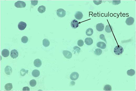
Reticulocytes are immature red blood cells, typically composing about 1% of the red cells in the human body. Reticulocytes develop and mature in the red bone marrow and then circulate for about a day in the blood stream before developing into mature red blood cells. Like mature red blood cells, reticulocytes do not have a cell nucleus. They are called reticulocytes because of a reticular (mesh-like) network of ribosomal RNA that becomes visible under a microscope with certain stains such as new methylene blue.
Differential Diagnosis of Reticulocytosis
| Cardiovascular | No underlying causes |
| Chemical / poisoning | No underlying causes |
| Dermatologic | No underlying causes |
| Drug Side Effect | No underlying causes |
| Ear Nose Throat | No underlying causes |
| Endocrine | No underlying causes |
| Environmental | No underlying causes |
| Gastroenterologic | No underlying causes |
| Genetic | No underlying causes |
| Hematologic | Acute Myelogenous/Blastic Leukemia AML, Leukemia, Lymphoma/malignant, Myeloproliferative disease, non-Hodgkins lymphoma, Primary Myelofibrosis/Myeloid metaplasia, Folate depletion, Folic acid deficiency anemia, Iron deficiency anemia, Pellagra/niacin deficiency, Vitamin B12 deficiency |
| Iatrogenic | No underlying causes |
| Infectious Disease | Abscesses, Bacteremia/Septicemia, Infected organ, Parvovirus Infection/Parvovirus B19, Posthepatitic aplastic anemia, Tuberculosis of bone marrow |
| Musculoskeletal / Ortho | No underlying causes |
| Neurologic | No underlying causes |
| Nutritional / Metabolic | Impaired folic acid metabolism, Anemia of malnutrition, Folate depletion, Folic acid deficiency anemia, Inadequate Folic acid in diet, Iron deficiency anemia, Iron deficient diet, Malnutrition/Starvation, Folic acid dependency/metabolic defect, Kwashiorkor (protein deficiency,severe), Malabsorption of folic acid, Pellagra/niacin deficiency, Vitamin B12 deficiency |
| Obstetric/Gynecologic | No underlying causes |
| Oncologic | Acute Myelogenous/Blastic Leukemia AML, Leukemia, Lymphoma/malignant, Myeloproliferative disease, non-Hodgkins lymphoma, Primary Myelofibrosis/Myeloid metaplasia |
| Opthalmologic | No underlying causes |
| Overdose / Toxicity | No underlying causes |
| Psychiatric | No underlying causes |
| Pulmonary | No underlying causes |
| Renal / Electrolyte | No underlying causes |
| Rheum / Immune / Allergy | No underlying causes |
| Sexual | No underlying causes |
| Trauma | No underlying causes |
| Urologic | No underlying causes |
| Miscellaneous | No underlying causes |
Differential Diagnosis of Reticulocytosis
(In alphabetical order)
- Acquired sideroblastic anemia
- Addison's disease (chronic adrenal insufficiency)
- Adrenocorticoid (Isolated) Deficiency
- AML (AML)
- Androgen insensitivity syndrome
- Anemia of chronic disease
- Anemia of malnutrition
- Anemia of uremia
- Antimetabolite medication Administration/Toxicity
- Aplastic anemia
- Aplastic anemia crisis
- Bacteremia/Septicemia
- Chemotherapy, cancer (anti-neoplastic)
- Chronic alcoholism
- Chronic liver disease
- Chronic renal failure
- Combined system disease/pernicious anemia
- Congenital aplastic anemia
- Congenital folate malabsorption
- Drug induced anemia
- Drug induced Bone marrow suppression.
- Fanconi's pancytopenia-dysmelia syndrome
- Folate depletion
- Folic acid deficiency anemia
- Folic acid dependency/metabolic defect
- Hemoglobin H disease
- Hereditary elliptocytosis
- Hereditary sideroblastic anemia
- Hypogonadism,
- Hypothyroidism (myxedema)
- Impaired folic acid metabolism
- Inadequate Folic acid in diet
- Infected organ, Abscesses
- Iron deficiency anemia
- Iron deficient diet
- Klinefelter's syndrome
- Kwashiorkor (severe protein deficiency)
- Lead poisoning
- Leukemia
- Liver disease/Liver disorders
- Malabsorption of folic acid
- Malnutrition/Starvation
- Megaloblastic anemia
- Megaloblastic anemia of pregnancy
- Methotrexate (Rheumatrex) Administration/Toxicity
- Myeloid metaplasia pathophysiology
- Myelophthisic anemia
- Myeloproliferative disease
- Non-Hodgkins lymphoma/malignant,
- Parvovirus Infection/Parvovirus B19
- Pellagra/niacin deficiency
- Pernicious anemia
- Posthepatitic aplastic anemia
- Postirradiation effects
- Primary Myelofibrosis/Myeloid metaplasia
- Radiation exposure
- Refractory megaloblastic anemia
- Secondary myelofibrosis
- Severe, acute Radiation sickness
- Sideroblastic Anemia Siderochrestic
- Tetraethyl lead poisoning
- Thalassemia major
- Thalassemia minor
- Thalassemia-hemoglobin C disease
- Tuberculosis of bone marrow
- Vitamin B12 deficiency
Diagnosis
Laboratory Findings
Reticulocytes appear slightly bluer than other red cells when looked at with the normal Romanowsky stain. Reticulocytes are also slightly larger, which can be picked up as a high MCV (mean corpuscular volume) with a full blood count done by a trained medical scientist, who has specialized in haematology, or a machine. [1] [2] [3]
The reticulocyte count is the percentage of circulating red blood cells that are in the reticulocyte stage.
To accurately measure reticulocyte counts, automated counters that use lasers mark cell samples with fluorescent dye that marks RNA and DNA (such as thiazole orange).[4] This distinguishes reticulocytes as the middle ground of dye response to laser light, between red blood cells (which have neither RNA nor DNA) and lymphocytes (which have a large amount of DNA, unlike reticulocytes).[5]
The normal range of values for reticulocytes in the blood depends on the clinical situation and the lab, but broadly speaking is 0.5% to 1.5%. However, if a person has anaemia, their reticulocyte percentage should be higher than "normal" if the bone marrow's ability to produce new blood cells remains intact. Thus, calculating the reticulocyte production index is an important step in understanding whether the reticulocyte count is appropriate or inappropriate to the situation. This is often a more important question than whether the percentage is in the normal range; for instance, if someone is anemic but only has a reticulocyte percentage of 1%, this means that the bone marrow is likely not producing new blood cells at a rate that will correct the anemia. The number of reticulocytes is a good indicator of bone marrow activity, because it represents recent production. This means that the reticulocyte count, and the reticulocyte production index that can be calculated from it, can be used to determine whether a production problem is contributing to the anaemia, and can also be used to monitor the progress of treatment for anaemia.
The specimen requirement for a reticulocyte count is EDTA anti-coagulated whole blood (lavender-top bottle if using the Vacutainer®, Vacuette® or Monoject® systems; red-top if using the S-Monovette® system).
When there is an increased production of red blood cells to overcome chronic or severe loss of mature red blood cells, such as in a haemolytic anaemia, people often have a markedly high number and percentage of reticulocytes. A very high number of reticulocytes in the blood can be described as reticulocytosis.
Abnormally low numbers of reticulocytes can be attributed to chemotherapy, aplastic anaemia, pernicious anaemia, bone marrow malignancies, problems of erythropoietin production, or other causes of anaemia due to poor RBC production.
-
Reticulocyte (Courtesy of Melih Aktan M.D.)
-
Reticulocyte (Courtesy of Melih Aktan M.D.)


-

Reticulocyte
-

Erythrocyte


See also
References
- ↑ The Disease Database
- ↑ Sailer, Christian, Wasner, Susanne. Differential Diagnosis Pocket. Hermosa Beach, CA: Borm Bruckmeir Publishing LLC, 2002:77 ISBN 1591032016
- ↑ Kahan, Scott, Smith, Ellen G. In A Page: Signs and Symptoms. Malden, Massachusetts: Blackwell Publishing, 2004:68 ISBN 140510368X
- ↑ Davis BH, Bigelow NC (1994). "Reticulocyte analysis and reticulocute maturity index". In Darzynkiewicz Z, Crissman HA (eds.). Flow cytometry. Methods in Cell Biology. 42. San Diego: Academic Press. pp. 263&ndash, 74. ISBN 0-12203-052-4.
- ↑ http://www.medicaldesign.com/articles/ID/532
Template:Blood Template:SIB he:רטיקולוציט id:Retikulosit fi:Retikulosyytti sq:Retikulociti