Dextro-transposition of the great arteries chest X ray: Difference between revisions
Jump to navigation
Jump to search
No edit summary |
No edit summary |
||
| Line 26: | Line 26: | ||
*[http://www.kumc.edu/instruction/medicine/pedcard/cardiology/pedcardio/dtgadiagram.gif Diagram at kumc.edu] | *[http://www.kumc.edu/instruction/medicine/pedcard/cardiology/pedcardio/dtgadiagram.gif Diagram at kumc.edu] | ||
*[http://www.med.umich.edu/cvc/mchc/partran.htm Diagram and description at umich.edu] | *[http://www.med.umich.edu/cvc/mchc/partran.htm Diagram and description at umich.edu] | ||
*[http://www.rch.org.au/cardiology/defects.cfm?doc_id=5098 Royal Children's Hospital, Melbourne] | *[http://www.rch.org.au/cardiology/defects.cfm?doc_id=5098 Royal Children's Hospital, Melbourne] | ||
*[http://www.mayoclinic.org/corrected-transposition-great-arteries Mayo Clinic, Arizona - Florida - Minnesota, USA] | *[http://www.mayoclinic.org/corrected-transposition-great-arteries Mayo Clinic, Arizona - Florida - Minnesota, USA] | ||
Revision as of 04:16, 14 August 2011
|
Dextro-transposition of the great arteries/complete transposition of the great arteries Microchapters |
|
Differentiating dextro-transposition of the great arteries from other Diseases |
|---|
|
Diagnosis |
Editor-In-Chief: C. Michael Gibson, M.S., M.D. [1]; Associate Editor(s)-In-Chief: Priyamvada Singh, M.B.B.S. [2]; Cafer Zorkun, M.D., Ph.D. [3]; Keri Shafer, M.D. [4]; Assistant Editor(s)-In-Chief: Kristin Feeney, B.S. [5]
Overview
Chest Xray may show the classical egg-on-side pattern
Chest X Ray
- Generally, the superior mediastinum may be narrow due to the anterior-posterior relationship of the great vessels.
- Initially, cardiac size is normal, but soon enlarges with the cardiac apex shifted to the left and inferiorly, producing the typically ovale-shaped or egg-on-side pattern.
- If a VSD is present, there will be an increase of the pulmonar vascular margins.
-
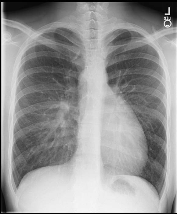
Transposition of great vessels.
-

Transposition of great vessels.


References
Acknowledgements and Initial Contributors to Page
Leida Perez, M.D.
External links
- Diagram at kumc.edu
- Diagram and description at umich.edu
- Royal Children's Hospital, Melbourne
- Mayo Clinic, Arizona - Florida - Minnesota, USA