Sickle-cell disease x ray: Difference between revisions
Jump to navigation
Jump to search
Created page with "{{Sickle-cell disease}} {{CMG}}; {{AE}} {{AN}} ==Overview== ==X ray findings== (Images shown below are courtesy of RadsWiki) '''Patient #1: SCD patient with H shaped verteb..." |
Shyam Patel (talk | contribs) No edit summary |
||
| Line 1: | Line 1: | ||
{{Sickle-cell disease}} | {{Sickle-cell disease}} | ||
{{CMG}}; {{AE}} {{AN}} | {{CMG}}; {{AE}} {{AN}} {{shyam}} | ||
==Overview== | ==Overview== | ||
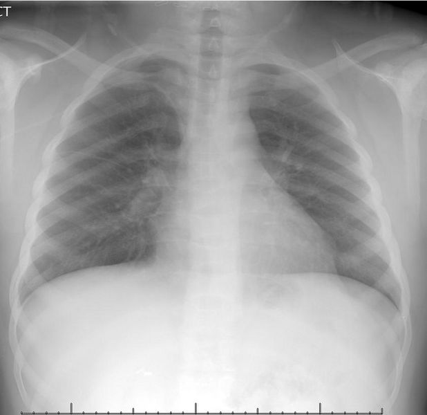
An X-ray can be useful if a patient presents with pulmonary symptoms, which can be due to pneumonia or acute chest syndrome. | |||
==X ray findings== | ==X ray findings== | ||
Revision as of 00:34, 29 August 2016
|
Sickle-cell disease Microchapters |
|
Diagnosis |
|---|
|
Treatment |
|
Case Studies |
|
Sickle-cell disease x ray On the Web |
|
American Roentgen Ray Society Images of Sickle-cell disease x ray |
|
Risk calculators and risk factors for Sickle-cell disease x ray |
Editor-In-Chief: C. Michael Gibson, M.S., M.D. [1]; Associate Editor(s)-in-Chief: Aarti Narayan, M.B.B.S [2] Shyam Patel [3]
Overview
An X-ray can be useful if a patient presents with pulmonary symptoms, which can be due to pneumonia or acute chest syndrome.
X ray findings
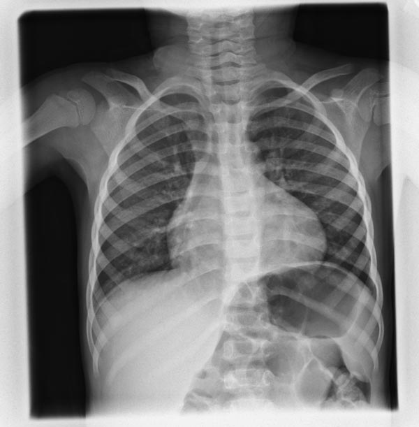
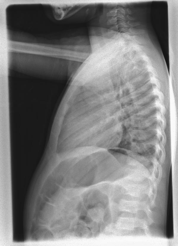
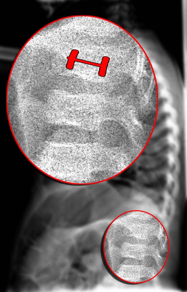
(Images shown below are courtesy of RadsWiki)
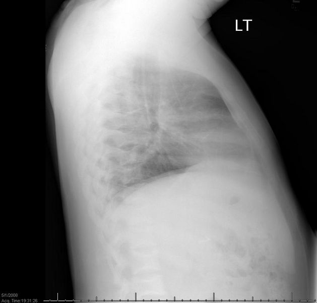
Patient #1: SCD patient with H shaped vertebrae
Patient #1: SCD patient with H shaped vertebrae
