Chancroid physical examination: Difference between revisions
No edit summary |
No edit summary |
||
| Line 24: | Line 24: | ||
*Males typically have a single ulcer | *Males typically have a single ulcer | ||
*Females typically have multiplee ulcers | *Females typically have multiplee ulcers | ||
*May look similar to [[Syphilis|syphilitic]] [[chancre]] | |||
===Common locations in males=== | ===Common locations in males=== | ||
| Line 39: | Line 40: | ||
*Inner thighs (least common) | *Inner thighs (least common) | ||
===Other Signs=== | |||
*Inguinal lymphadenitis in approximately 50% of patients, known as [[Bubo|buboes]] (typically unilateral and predominantly in males)<ref name="Lewis2003">{{cite journal|last1=Lewis|first1=D A|title=Chancroid: clinical manifestations, diagnosis, and management|journal=Sexually Transmitted Infections|volume=79|issue=1|year=2003|pages=68–71|issn=13684973|doi=10.1136/sti.79.1.68}}</ref> | |||
*Approximately 25% of patients with lymphadenopathy may present with abscesses in lymph nodes or ruptured buboes | |||
==Gallery== | |||
<gallery> | <gallery> | ||
Image:Chancroid01.jpg|Chancroid. <SMALL><SMALL>''[http://www.atlasdermatologico.com.br/ Adapted from Dermatology Atlas.]''<ref name="Dermatology Atlas">{{Cite web | title = Dermatology Atlas | url = http://www.atlasdermatologico.com.br/}}</ref></SMALL></SMALL> | Image:Chancroid01.jpg|Chancroid. <SMALL><SMALL>''[http://www.atlasdermatologico.com.br/ Adapted from Dermatology Atlas.]''<ref name="Dermatology Atlas">{{Cite web | title = Dermatology Atlas | url = http://www.atlasdermatologico.com.br/}}</ref></SMALL></SMALL> | ||
Image:Chancroid02.jpg|Chancroid. <SMALL><SMALL>''[http://www.atlasdermatologico.com.br/ Adapted from Dermatology Atlas.]''<ref name="Dermatology Atlas">{{Cite web | title = Dermatology Atlas | url = http://www.atlasdermatologico.com.br/}}</ref></SMALL></SMALL> | Image:Chancroid02.jpg|Chancroid. <SMALL><SMALL>''[http://www.atlasdermatologico.com.br/ Adapted from Dermatology Atlas.]''<ref name="Dermatology Atlas">{{Cite web | title = Dermatology Atlas | url = http://www.atlasdermatologico.com.br/}}</ref></SMALL></SMALL> | ||
Revision as of 20:56, 21 January 2016
|
Chancroid Microchapters |
|
Diagnosis |
|---|
|
Treatment |
|
Case Studies |
|
Chancroid physical examination On the Web |
|
American Roentgen Ray Society Images of Chancroid physical examination |
|
calculators and risk factors for Chancroid physical examination |
Editor-In-Chief: C. Michael Gibson, M.S., M.D. [1]; Associate Editor(s)-in-Chief: Yazan Daaboul, M.D. Nate Michalak, B.A. Serge Korjian M.D.
Overview
Physical Examination
Vital Signs
Typically normal
Skin
A patient may present with either of the following types of lesions on the genitals, depending on the stage of infection:[1][2]
Ulcer characteristics:[3]
- Ranges in size from 3 to 50 mm (1/8 to 2 inches) in diameter
- Painful
- Soft, nonindurated
- Irregular border
- Sharp margins
- Grey/yellow exudate
- Males typically have a single ulcer
- Females typically have multiplee ulcers
- May look similar to syphilitic chancre
Common locations in males
- Foreskin (prepuce) (most common)
- Groove behind the head of the penis (coronal sulcus)
- Shaft of the penis
- Head of the penis (glans penis)
- Opening of the penis (urethral meatus)
- Scrotum (least common)
Common locations in females
- labia majora (most common). "Kissing ulcers" may develop, defined as ulcers that occur on opposing surfaces of the labia.
- labia minora
- Perineal area
- Inner thighs (least common)
Other Signs
- Inguinal lymphadenitis in approximately 50% of patients, known as buboes (typically unilateral and predominantly in males)[4]
- Approximately 25% of patients with lymphadenopathy may present with abscesses in lymph nodes or ruptured buboes
Gallery
-
![Chancroid. Adapted from Dermatology Atlas.[5]](/images/6/6f/Chancroid01.jpg)
Chancroid. Adapted from Dermatology Atlas.[5]
-
![Chancroid. Adapted from Dermatology Atlas.[5]](/images/c/c1/Chancroid02.jpg)
Chancroid. Adapted from Dermatology Atlas.[5]
-
![Chancroid. Adapted from Dermatology Atlas.[5]](/images/1/1a/Chancroid03.jpg)
Chancroid. Adapted from Dermatology Atlas.[5]
-
![Chancroid. Adapted from Dermatology Atlas.[5]](/images/7/70/Chancroid04.jpg)
Chancroid. Adapted from Dermatology Atlas.[5]
-
![Chancroid. Adapted from Dermatology Atlas.[5]](/images/2/22/Chancroid05.jpg)
Chancroid. Adapted from Dermatology Atlas.[5]
-
![Chancroid. Adapted from Dermatology Atlas.[5]](/images/d/d6/Chancroid06.jpg)
Chancroid. Adapted from Dermatology Atlas.[5]
-
![Chancroid. Adapted from Dermatology Atlas.[5]](/images/1/19/Chancroid07.jpg)
Chancroid. Adapted from Dermatology Atlas.[5]
-
![Chancroid. Adapted from Dermatology Atlas.[5]](/images/6/60/Chancroid08.jpg)
Chancroid. Adapted from Dermatology Atlas.[5]
-
![Chancroid. Adapted from Dermatology Atlas.[5]](/images/4/4b/Chancroid09.jpg)
Chancroid. Adapted from Dermatology Atlas.[5]
-
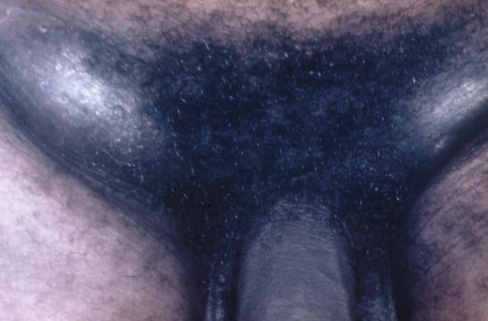
Chancroid infection has spread to the inguinal lymph nodes, which have enlarged forming buboes.
-
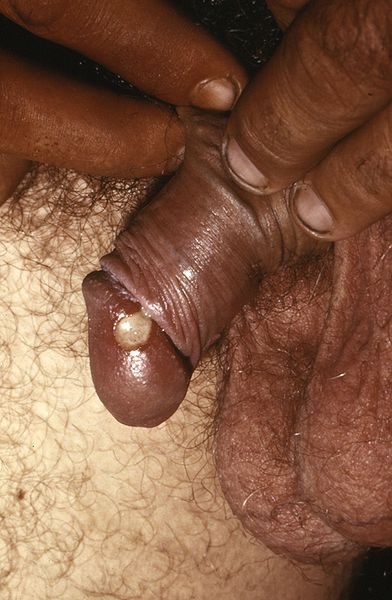
Penile chancroid lesion.
![Chancroid. Adapted from Dermatology Atlas.[5]](/index.php/File:Chancroid01.jpg)
![Chancroid. Adapted from Dermatology Atlas.[5]](/index.php/File:Chancroid02.jpg)
![Chancroid. Adapted from Dermatology Atlas.[5]](/index.php/File:Chancroid03.jpg)
![Chancroid. Adapted from Dermatology Atlas.[5]](/index.php/File:Chancroid04.jpg)
![Chancroid. Adapted from Dermatology Atlas.[5]](/index.php/File:Chancroid05.jpg)
![Chancroid. Adapted from Dermatology Atlas.[5]](/index.php/File:Chancroid06.jpg)
![Chancroid. Adapted from Dermatology Atlas.[5]](/index.php/File:Chancroid07.jpg)
![Chancroid. Adapted from Dermatology Atlas.[5]](/index.php/File:Chancroid08.jpg)
![Chancroid. Adapted from Dermatology Atlas.[5]](/index.php/File:Chancroid09.jpg)


References
- ↑ Chancroid. UpToDate (September 25, 2015). http://www.uptodate.com/contents/chancroid#H3 Accessed January 19, 2016.
- ↑ Spinola, S. M. (2002). "Immunopathogenesis of Haemophilus ducreyi Infection (Chancroid)". Infection and Immunity. 70 (4): 1667–1676. doi:10.1128/IAI.70.4.1667-1676.2002. ISSN 0019-9567.
- ↑ Chancroid. Wikipedia (July 16, 2015). https://en.wikipedia.org/wiki/Chancroid Accessed January 15, 2016.
- ↑ Lewis, D A (2003). "Chancroid: clinical manifestations, diagnosis, and management". Sexually Transmitted Infections. 79 (1): 68–71. doi:10.1136/sti.79.1.68. ISSN 1368-4973.
- ↑ 5.0 5.1 5.2 5.3 5.4 5.5 5.6 5.7 5.8 "Dermatology Atlas".