Oligodendroglioma pathophysiology: Difference between revisions
No edit summary |
|||
| Line 39: | Line 39: | ||
:*[[Posterior fossa]] (rare) | :*[[Posterior fossa]] (rare) | ||
:*[[spinal cord|Intramedullary spinal cord]] (very rare) | :*[[spinal cord|Intramedullary spinal cord]] (very rare) | ||
====Gallery==== | |||
<gallery> | |||
Image:Glioma Gross 4.jpg|Brain: Oligodendroglioma: Gross; natural color, large, well circumscribed lesion in left [[frontal lobe]] | |||
Image:Oligodendroglioma gross 2.jpg|They arise from glial cells called oligodendrocytes.<ref name=gvuhjgj>Image of oligodendroglioma. http://drarunlnaik.com/oligodendroglioma/</ref> | |||
===Microscopic Pathology=== | ===Microscopic Pathology=== | ||
| Line 67: | Line 73: | ||
*[[vascular|Microvacular proliferation]] | *[[vascular|Microvacular proliferation]] | ||
**Either in the form of 'glomeruloid' vessels or endothelial [[hyperplasia]] | **Either in the form of 'glomeruloid' vessels or endothelial [[hyperplasia]] | ||
====Gallery==== | |||
<gallery> | |||
Image:Oligodendroglioma1 low mag.jpg|Oligodendroglioma low magnification showing the characteristic small, branching, ''chicken wire''-like blood vessels.H&E stain.<ref name=microscope>Images of microscopic appearance of oligodendroglioma. Wikipedia 2015. https://en.wikipedia.org/wiki/Oligodendroglioma</ref> | |||
Image:Oligodendroglioma1 high mag.jpg|Oligodendroglioma high magnification showing highly cellular lesion composed of cells resembling ''fried eggs'' with distinct cell borders moderate-to-marked nuclear atypia, and a clear cytoplasm. Acutely branched capillary sized vessels - "''chicken-wire''" like appearance.<ref name=microscope>Images of microscopic appearance of oligodendroglioma. Wikipedia 2015. https://en.wikipedia.org/wiki/Oligodendroglioma</ref> | |||
Image:Oligodendroglioma discrete invasion HE.jpg|Low power magnification of oligodendroglioma biopsy specimen showing discrete infiltration of the surrounding brain (HE stain, x40 mag).<ref name=LP>Images of oligodendroglioma. Libre Pathology 2015. http://librepathology.org/wiki/index.php/Oligodendroglioma</ref> | |||
Image:Anaplastic oligodendroglioma minigemistocytes.jpg|Histopathology of anaplastic oligodendroglioma showing minigemistocytes and mitoses among tumor cells with perinuclear halo. HE stain.<ref name=LP>Images of oligodendroglioma. Libre Pathology 2015. http://librepathology.org/wiki/index.php/Oligodendroglioma</ref> | |||
Image:MAP2 anaplastic oligodendroglioma.jpg|Histopathology of anaplastic oligodendroglioma (MAP2 staining) showing perinuclear immunoreactivity of tumor cells.<ref name=LP>Images of oligodendroglioma. Libre Pathology 2015. http://librepathology.org/wiki/index.php/Oligodendroglioma</ref> | |||
Image:IDH1 R132H in anaplastic ologodendroglioma.jpg|Histopathology of anaplastic oligodendroglioma (IDH1-R132H staining) showing immunoreactivity of tumor cells indicating presence of the isocitrate dehydrogenase 1-R132H mutation.<ref name=LP>Images of oligodendroglioma. Libre Pathology 2015. http://librepathology.org/wiki/index.php/Oligodendroglioma</ref> | |||
</gallery> | |||
===Immunohistochemistry=== | ===Immunohistochemistry=== | ||
| Line 82: | Line 98: | ||
*[[OLIG1]] | *[[OLIG1]] | ||
*[[OLIG2]] | *[[OLIG2]] | ||
==References== | ==References== | ||
Revision as of 19:37, 13 October 2015
Editor-In-Chief: C. Michael Gibson, M.S., M.D. [1]Associate Editor(s)-in-Chief: Sujit Routray, M.D. [2]
|
Oligodendroglioma Microchapters |
|
Diagnosis |
|---|
|
Treatment |
|
Case Studies |
|
Oligodendroglioma pathophysiology On the Web |
|
American Roentgen Ray Society Images of Oligodendroglioma pathophysiology |
|
Risk calculators and risk factors for Oligodendroglioma pathophysiology |
Overview
Pathophysiology
Pathogenesis
- Oligodendroglioma does not arise from the bipotential oligodendrocytes, although tumor cells look very similiar.[1]
- Oligodendroglioma arises from the tripotential glial precursor cells.
Genetics
- Development of oligodendroglioma is the result from multiple genetic mutations.
- Genes associated with the pathogenesis of oligodendroglioma include:[2][3][4][5][6][7][8][9][10]
- There is a strong association of oligodendroglioma with expression of receptor tyrosine kinases that activate PI3K/AKT, RAS/MAP, and PLC/PKC pathways.[10]
Gross Pathology
- On gross pathology, oligodendroglioma is characterized by a well-circumscribed, gelatinous, gray mass which may expand a gyrus and remodel the skull.[11]
- Other characteristic gross pathological features associated with oligodendroglioma include:[11][10]
- Calcification (70-90%; one of the most frequently calcifying tumors)
- Focal hemorrhage
- Cystic (20%)
- Common intracranial sites associated with oligodendroglioma include:[12]
- Cerebral hemispheres - distribution between frontal, parietal, temporal, and occipital lobe approximates 3:2:2:1
- Posterior fossa (rare)
- Intramedullary spinal cord (very rare)
Gallery
-
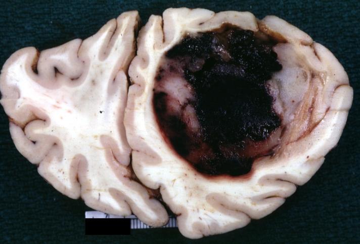
Brain: Oligodendroglioma: Gross; natural color, large, well circumscribed lesion in left frontal lobe
-
![They arise from glial cells called oligodendrocytes.[13]](/images/b/b6/Oligodendroglioma_gross_2.jpg)
They arise from glial cells called oligodendrocytes.[13]
-
-
-
-
-
-
-
-
-
![Oligodendroglioma low magnification showing the characteristic small, branching, chicken wire-like blood vessels.H&E stain.[14]](/images/7/7c/Oligodendroglioma1_low_mag.jpg)
Oligodendroglioma low magnification showing the characteristic small, branching, chicken wire-like blood vessels.H&E stain.[14]
-
![Oligodendroglioma high magnification showing highly cellular lesion composed of cells resembling fried eggs with distinct cell borders moderate-to-marked nuclear atypia, and a clear cytoplasm. Acutely branched capillary sized vessels - "chicken-wire" like appearance.[14]](/images/a/a4/Oligodendroglioma1_high_mag.jpg)
Oligodendroglioma high magnification showing highly cellular lesion composed of cells resembling fried eggs with distinct cell borders moderate-to-marked nuclear atypia, and a clear cytoplasm. Acutely branched capillary sized vessels - "chicken-wire" like appearance.[14]
-
![Low power magnification of oligodendroglioma biopsy specimen showing discrete infiltration of the surrounding brain (HE stain, x40 mag).[15]](/images/3/39/Oligodendroglioma_discrete_invasion_HE.jpg)
Low power magnification of oligodendroglioma biopsy specimen showing discrete infiltration of the surrounding brain (HE stain, x40 mag).[15]
-
![Histopathology of anaplastic oligodendroglioma showing minigemistocytes and mitoses among tumor cells with perinuclear halo. HE stain.[15]](/images/5/54/Anaplastic_oligodendroglioma_minigemistocytes.jpg)
Histopathology of anaplastic oligodendroglioma showing minigemistocytes and mitoses among tumor cells with perinuclear halo. HE stain.[15]
-
![Histopathology of anaplastic oligodendroglioma (MAP2 staining) showing perinuclear immunoreactivity of tumor cells.[15]](/images/1/16/MAP2_anaplastic_oligodendroglioma.jpg)
Histopathology of anaplastic oligodendroglioma (MAP2 staining) showing perinuclear immunoreactivity of tumor cells.[15]
-
![Histopathology of anaplastic oligodendroglioma (IDH1-R132H staining) showing immunoreactivity of tumor cells indicating presence of the isocitrate dehydrogenase 1-R132H mutation.[15]](/images/1/1c/IDH1_R132H_in_anaplastic_ologodendroglioma.jpg)
Histopathology of anaplastic oligodendroglioma (IDH1-R132H staining) showing immunoreactivity of tumor cells indicating presence of the isocitrate dehydrogenase 1-R132H mutation.[15]

![They arise from glial cells called oligodendrocytes.[13]](/index.php/File:Oligodendroglioma_gross_2.jpg)
![Oligodendroglioma low magnification showing the characteristic small, branching, chicken wire-like blood vessels.H&E stain.[14]](/index.php/File:Oligodendroglioma1_low_mag.jpg)
![Oligodendroglioma high magnification showing highly cellular lesion composed of cells resembling fried eggs with distinct cell borders moderate-to-marked nuclear atypia, and a clear cytoplasm. Acutely branched capillary sized vessels - "chicken-wire" like appearance.[14]](/index.php/File:Oligodendroglioma1_high_mag.jpg)
![Low power magnification of oligodendroglioma biopsy specimen showing discrete infiltration of the surrounding brain (HE stain, x40 mag).[15]](/index.php/File:Oligodendroglioma_discrete_invasion_HE.jpg)
![Histopathology of anaplastic oligodendroglioma showing minigemistocytes and mitoses among tumor cells with perinuclear halo. HE stain.[15]](/index.php/File:Anaplastic_oligodendroglioma_minigemistocytes.jpg)
![Histopathology of anaplastic oligodendroglioma (MAP2 staining) showing perinuclear immunoreactivity of tumor cells.[15]](/index.php/File:MAP2_anaplastic_oligodendroglioma.jpg)
![Histopathology of anaplastic oligodendroglioma (IDH1-R132H staining) showing immunoreactivity of tumor cells indicating presence of the isocitrate dehydrogenase 1-R132H mutation.[15]](/index.php/File:IDH1_R132H_in_anaplastic_ologodendroglioma.jpg)
Immunohistochemistry
Oligodendroglioma is demonstrated by positivity to tumor markers such as:[16][17][10]
- MAP2
- GFAP
- S-100
- EMA
- IDH1-R132H
- ATRX
- Ki-67
- NSE
- Synaptophysin
- OLIG1
- OLIG2
References
- ↑ General features of oligodendroglioma. Libre Pathology. http://librepathology.org/wiki/index.php/Oligodendroglioma#cite_note-1
- ↑ Molecular genetics of oligodendroglioma. https://en.wikipedia.org/wiki/Oligodendroglioma
- ↑ Bettegowda C, Agrawal N, Jiao Y, Sausen M, Wood LD, Hruban RH; et al. (2011). "Mutations in CIC and FUBP1 contribute to human oligodendroglioma". Science. 333 (6048): 1453–5. doi:10.1126/science.1210557. PMC 3170506. PMID 21817013.
- ↑ Prognosis and treatment of oligodendroglioma. Wikipedia 2015. https://en.wikipedia.org/wiki/Oligodendroglioma
- ↑ Yip S, Butterfield YS, Morozova O, Chittaranjan S, Blough MD, An J; et al. (2012). "Concurrent CIC mutations, IDH mutations, and 1p/19q loss distinguish oligodendrogliomas from other cancers". J Pathol. 226 (1): 7–16. doi:10.1002/path.2995. PMC 3246739. PMID 22072542.
- ↑ Labreche K, Simeonova I, Kamoun A, Gleize V, Chubb D, Letouzé E; et al. (2015). "TCF12 is mutated in anaplastic oligodendroglioma". Nat Commun. 6: 7207. doi:10.1038/ncomms8207. PMC 4490400. PMID 26068201.
- ↑ Suri V, Jha P, Agarwal S, Pathak P, Sharma MC, Sharma V; et al. (2011). "Molecular profile of oligodendrogliomas in young patients". Neuro Oncol. 13 (10): 1099–106. doi:10.1093/neuonc/nor146. PMC 3177666. PMID 21937591.
- ↑ Hagel C, Laking G, Laas R, Scheil S, Jung R, Milde-Langosch K; et al. (1996). "Demonstration of p53 protein and TP53 gene mutations in oligodendrogliomas". Eur J Cancer. 32A (13): 2242–8. PMID 9038605.
- ↑ Schmoldt A, Benthe HF, Haberland G (1975). "Digitoxin metabolism by rat liver microsomes". Biochem Pharmacol. 24 (17): 1639–41. PMID doi:10.1016/S0090-3019(03)00167-8 Check
|pmid=value (help). - ↑ 10.0 10.1 10.2 10.3 von Deimling, A; Hartmann, C (2005). "Oligodendrogliomas: Impact of molecular genetics on treatment". Neurology India. 53 (2): 140. doi:10.4103/0028-3886.16394. ISSN 0028-3886.
- ↑ 11.0 11.1 Gross appearance of oligodendroglioma. Dr Henry Knipe and Dr Frank Gaillard et al. http://radiopaedia.org/articles/oligodendroglioma
- ↑ Gross/radiologic findings of oligodendroglioma. Libre Pathology. http://librepathology.org/wiki/index.php/Oligodendroglioma
- ↑ Image of oligodendroglioma. http://drarunlnaik.com/oligodendroglioma/
- ↑ 14.0 14.1 Images of microscopic appearance of oligodendroglioma. Wikipedia 2015. https://en.wikipedia.org/wiki/Oligodendroglioma
- ↑ 15.0 15.1 15.2 15.3 Images of oligodendroglioma. Libre Pathology 2015. http://librepathology.org/wiki/index.php/Oligodendroglioma
- ↑ IHC of oligodendroglioma. Libre Pathology. http://librepathology.org/wiki/index.php/Oligodendroglioma
- ↑ Hilbig A, Barbosa-Coutinho LM, Netto GC, Bleil CB, Toscani NV (2006). "[Immunohistochemistry in oligodendrogliomas]". Arq Neuropsiquiatr. 64 (1): 67–71. doi:/S0004-282X2006000100014 Check
|doi=value (help). PMID 16622556.