Aortic coarctation CT: Difference between revisions
m (Robot: Changing Category:DiseaseState to Category:Disease) |
No edit summary |
||
| (16 intermediate revisions by 5 users not shown) | |||
| Line 1: | Line 1: | ||
__NOTOC__ | |||
Please help WikiDoc by adding more content here. It's easy! Click [[Help:How_to_Edit_a_Page|here]] to learn about editing. | |||
{{Template:Aortic Coarctation}} | {{Template:Aortic Coarctation}} | ||
{{CMG}} | {{CMG}}; '''Associate Editor(s)-In-Chief:''' [[Priyamvada Singh|Priyamvada Singh, M.B.B.S.]][mailto:psingh13579@gmail.com], {{CZ}}; '''Assistant Editor(s)-In-Chief:''' [[Kristin Feeney|Kristin Feeney, B.S.]][mailto:kfeeney@elon.edu] | ||
==Overview== | |||
[[CT]] is useful in older or postoperative patients as it helps in to assessing residual [[obstruction]], [[hypoplasia]], [[aneurysms]], multiple surgical clips or a [[stent]] present in the area of coarctation <ref name="pmid8227813">{{cite journal |author=Mohiaddin RH, Kilner PJ, Rees S, Longmore DB |title=Magnetic resonance volume flow and jet velocity mapping in aortic coarctation |journal=[[Journal of the American College of Cardiology]] |volume=22 |issue=5 |pages=1515–21 |year=1993 |month=November |pmid=8227813 |doi= |url=http://linkinghub.elsevier.com/retrieve/pii/0735-1097(93)90565-I |accessdate=2012-04-14}}</ref>. | |||
==CT== | ==CT== | ||
Shown below are of CT scan images of aortic coarctation. | |||
{| | |||
|- | |||
| [[Image:Coarctation-of-the-aorta-CT-002.jpg|250px|Aortic Coarctation]] || [[Image:Coarctation-of-the-aorta-CT-003.jpg|Aortic Coarctation|250px]] | |||
|} | |||
<small>Courtesy of RadsWiki and copylefted</small> | |||
---- | |||
{| | |||
|- | |||
| [[Image:Coarctation-of-the-aorta-CT-004.jpg|Aortic Coarctation|250px]] || [[Image:Coarctation-of-the-aorta-CT-005.jpg|Aortic Coarctation|250px]] | |||
|} | |||
<small> Courtesy of Cafer Zorkun and copylefted </small> | |||
---- | |||
{| | |||
|- | |||
| [[Image:Coarctation-of-the-aorta-MRI-003.jpg|250px]] || [[Image:Coarctation-of-the-aorta-MRI-002.jpg|250px]] | |||
|} | |||
<small>Courtesy of RadsWiki and copylefted</small> | |||
---- | |||
<br> | |||
[[Image:Aortic coarctation MRI 001.jpg|250px]] | |||
<br clear'"left"/> | |||
<small> [http://www.peir.net Image courtesy of Professor Peter Anderson DVM PhD and published with permission © PEIR, University of Alabama at Birmingham, Department of Pathology] </small> | |||
== 2018 AHA/ACC Guideline for the Management of Adults With Congenital Heart Disease: A Report of the American College of Cardiology/American Heart Association Task Force on Clinical Practice Guidelines<ref name="pmid30121240">{{cite journal| author=Stout KK, Daniels CJ, Aboulhosn JA, Bozkurt B, Broberg CS, Colman JM | display-authors=etal| title=2018 AHA/ACC Guideline for the Management of Adults With Congenital Heart Disease: Executive Summary: A Report of the American College of Cardiology/American Heart Association Task Force on Clinical Practice Guidelines. | journal=J Am Coll Cardiol | year= 2019 | volume= 73 | issue= 12 | pages= 1494-1563 | pmid=30121240 | doi=10.1016/j.jacc.2018.08.1028 | pmc= | url=https://www.ncbi.nlm.nih.gov/entrez/eutils/elink.fcgi?dbfrom=pubmed&tool=sumsearch.org/cite&retmode=ref&cmd=prlinks&id=30121240 }}</ref> == | |||
=== Diagnostic Recommendations for Coarctation of the Aorta === | |||
{| class="wikitable" | |||
|- | |||
| colspan="1" style="text-align:center; background:LightGreen" |[[ACC AHA guidelines classification scheme#Classification of Recommendations|Class I]] | |||
|- | |||
| bgcolor="LightGreen" |'''1.''' Initial and follow-up aortic imaging using CMR or CTA is recommended in adults with coarctation of the aorta, including those who have had surgical or catheter intervention.''(Level of Evidence: B-NR)'' | |||
|} | |||
{| class="wikitable" | |||
|- | |||
| colspan="1" style="text-align:center; background:LemonChiffon" |[[ACC AHA guidelines classification scheme#Classification of Recommendations|Class IIb]] | |||
|- | |||
| bgcolor="LemonChiffon" |'''1.'''Screening for intracranial aneurysms by magnetic resonance angiography or CTA may be reasonable in adults with coarctation of the aorta.. ''(Level of Evidence: B-NR)'' | |||
|} | |||
==2008 ACC/AHA Guidelines for the Management of Adults With Congenital Heart Disease (DO NOT EDIT)<ref name="pmid19038677">{{cite journal| author=Warnes CA, Williams RG, Bashore TM, Child JS, Connolly HM, Dearani JA et al.| title=ACC/AHA 2008 guidelines for the management of adults with congenital heart disease: a report of the American College of Cardiology/American Heart Association Task Force on Practice Guidelines (Writing Committee to Develop Guidelines on the Management of Adults With Congenital Heart Disease). Developed in Collaboration With the American Society of Echocardiography, Heart Rhythm Society, International Society for Adult Congenital Heart Disease, Society for Cardiovascular Angiography and Interventions, and Society of Thoracic Surgeons. | journal=J Am Coll Cardiol | year= 2008 | volume= 52 | issue= 23 | pages= e1-121 | pmid=19038677 | doi=10.1016/j.jacc.2008.10.001 | pmc= | url=http://www.ncbi.nlm.nih.gov/entrez/eutils/elink.fcgi?dbfrom=pubmed&tool=sumsearch.org/cite&retmode=ref&cmd=prlinks&id=19038677 }} </ref> == | |||
=== Recommendations for Clinical Evaluation and Follow-Up (DO NOT EDIT)<ref name="pmid19038677">{{cite journal| author=Warnes CA, Williams RG, Bashore TM, Child JS, Connolly HM, Dearani JA et al.| title=ACC/AHA 2008 guidelines for the management of adults with congenital heart disease: a report of the American College of Cardiology/American Heart Association Task Force on Practice Guidelines (Writing Committee to Develop Guidelines on the Management of Adults With Congenital Heart Disease). Developed in Collaboration With the American Society of Echocardiography, Heart Rhythm Society, International Society for Adult Congenital Heart Disease, Society for Cardiovascular Angiography and Interventions, and Society of Thoracic Surgeons. | journal=J Am Coll Cardiol | year= 2008 | volume= 52 | issue= 23 | pages= e1-121 | pmid=19038677 | doi=10.1016/j.jacc.2008.10.001 | pmc= | url=http://www.ncbi.nlm.nih.gov/entrez/eutils/elink.fcgi?dbfrom=pubmed&tool=sumsearch.org/cite&retmode=ref&cmd=prlinks&id=19038677 }} </ref> === | |||
{|class="wikitable" | |||
|- | |||
| colspan="1" style="text-align:center; background:LightGreen"|[[ACC AHA guidelines classification scheme#Classification of Recommendations|Class I]] | |||
|- | |||
< | | bgcolor="LightGreen"|<nowiki>"</nowiki>'''1.''' Every patient with coarctation (repaired or not) should have at least one cardiovascular [[MRI]] or [[CT scan]] for complete evaluation of the [[thoracic aorta]] and intracranial vessels. ''([[ACC AHA guidelines classification scheme#Level of Evidence|Level of Evidence: B]])''<nowiki>"</nowiki> | ||
< | |||
</ | |||
|} | |||
==References== | ==References== | ||
{{reflist|2}} | {{reflist|2}} | ||
| Line 22: | Line 67: | ||
{{WS}} | {{WS}} | ||
[[CME Category::Cardiology]] | |||
[[Category:Cardiology]] | [[Category:Cardiology]] | ||
[[Category:Pediatrics]] | [[Category:Pediatrics]] | ||
[[Category:Disease]] | [[Category:Disease]] | ||
[[Category:Congenital heart disease]] | |||
[[Category:Needs content]] | |||
Latest revision as of 20:53, 15 December 2022
Please help WikiDoc by adding more content here. It's easy! Click here to learn about editing.
|
Aortic coarctation Microchapters |
|
Diagnosis |
|---|
|
Treatment |
|
Case Studies |
|
Aortic coarctation CT On the Web |
|
American Roentgen Ray Society Images of Aortic coarctation CT |
Editor-In-Chief: C. Michael Gibson, M.S., M.D. [1]; Associate Editor(s)-In-Chief: Priyamvada Singh, M.B.B.S.[2], Cafer Zorkun, M.D., Ph.D. [3]; Assistant Editor(s)-In-Chief: Kristin Feeney, B.S.[4]
Overview
CT is useful in older or postoperative patients as it helps in to assessing residual obstruction, hypoplasia, aneurysms, multiple surgical clips or a stent present in the area of coarctation [1].
CT
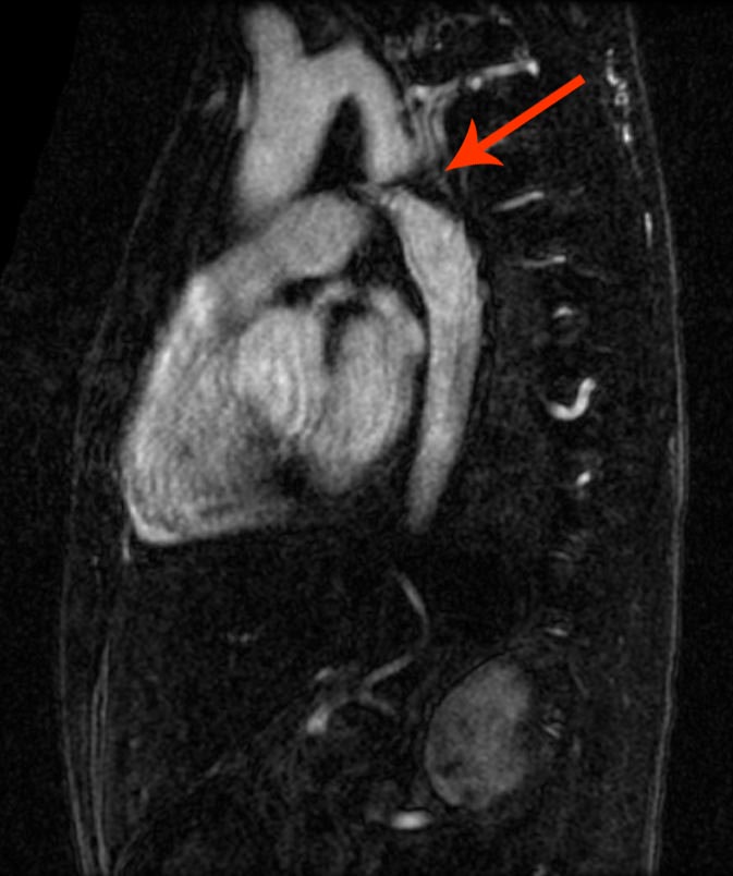
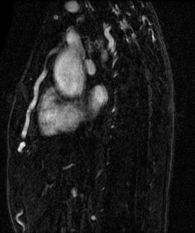
Shown below are of CT scan images of aortic coarctation.
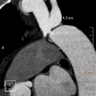 |
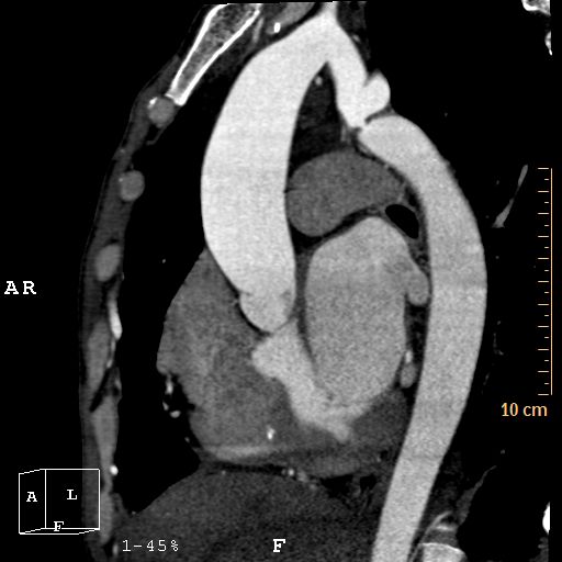
|
Courtesy of RadsWiki and copylefted
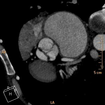 |
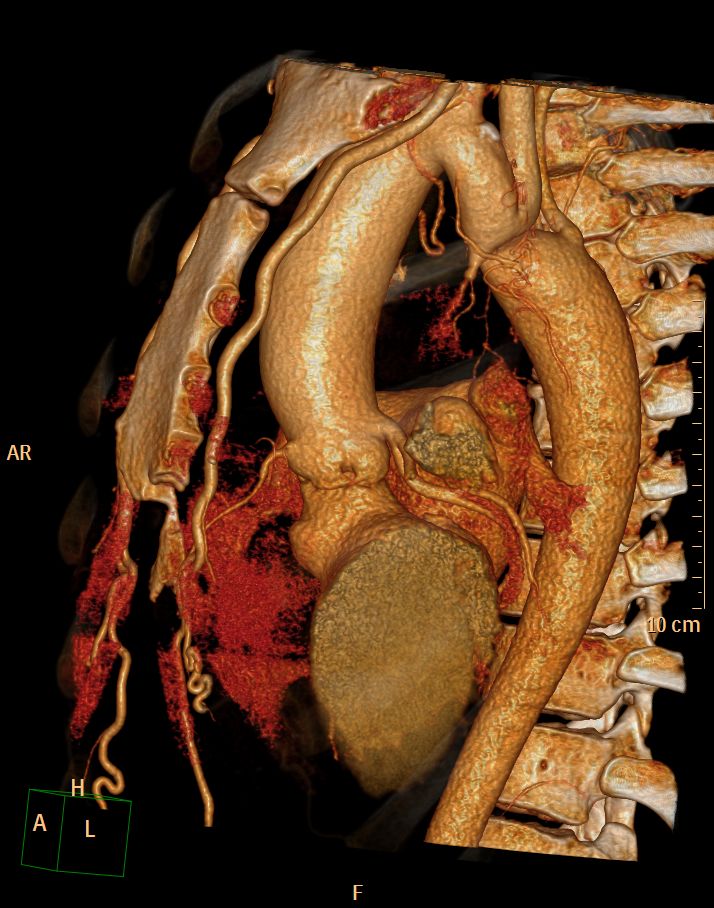
|
Courtesy of Cafer Zorkun and copylefted
 |
|
Courtesy of RadsWiki and copylefted
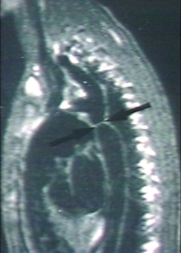
Image courtesy of Professor Peter Anderson DVM PhD and published with permission © PEIR, University of Alabama at Birmingham, Department of Pathology
2018 AHA/ACC Guideline for the Management of Adults With Congenital Heart Disease: A Report of the American College of Cardiology/American Heart Association Task Force on Clinical Practice Guidelines[2]
Diagnostic Recommendations for Coarctation of the Aorta
| Class I |
| 1. Initial and follow-up aortic imaging using CMR or CTA is recommended in adults with coarctation of the aorta, including those who have had surgical or catheter intervention.(Level of Evidence: B-NR) |
| Class IIb |
| 1.Screening for intracranial aneurysms by magnetic resonance angiography or CTA may be reasonable in adults with coarctation of the aorta.. (Level of Evidence: B-NR) |
2008 ACC/AHA Guidelines for the Management of Adults With Congenital Heart Disease (DO NOT EDIT)[3]
Recommendations for Clinical Evaluation and Follow-Up (DO NOT EDIT)[3]
| Class I |
| "1. Every patient with coarctation (repaired or not) should have at least one cardiovascular MRI or CT scan for complete evaluation of the thoracic aorta and intracranial vessels. (Level of Evidence: B)" |
References
- ↑ Mohiaddin RH, Kilner PJ, Rees S, Longmore DB (1993). "Magnetic resonance volume flow and jet velocity mapping in aortic coarctation". Journal of the American College of Cardiology. 22 (5): 1515–21. PMID 8227813. Retrieved 2012-04-14. Unknown parameter
|month=ignored (help) - ↑ Stout KK, Daniels CJ, Aboulhosn JA, Bozkurt B, Broberg CS, Colman JM; et al. (2019). "2018 AHA/ACC Guideline for the Management of Adults With Congenital Heart Disease: Executive Summary: A Report of the American College of Cardiology/American Heart Association Task Force on Clinical Practice Guidelines". J Am Coll Cardiol. 73 (12): 1494–1563. doi:10.1016/j.jacc.2018.08.1028. PMID 30121240.
- ↑ 3.0 3.1 Warnes CA, Williams RG, Bashore TM, Child JS, Connolly HM, Dearani JA; et al. (2008). "ACC/AHA 2008 guidelines for the management of adults with congenital heart disease: a report of the American College of Cardiology/American Heart Association Task Force on Practice Guidelines (Writing Committee to Develop Guidelines on the Management of Adults With Congenital Heart Disease). Developed in Collaboration With the American Society of Echocardiography, Heart Rhythm Society, International Society for Adult Congenital Heart Disease, Society for Cardiovascular Angiography and Interventions, and Society of Thoracic Surgeons". J Am Coll Cardiol. 52 (23): e1–121. doi:10.1016/j.jacc.2008.10.001. PMID 19038677.