Dextro-transposition of the great arteries chest X ray: Difference between revisions
(New page: {{Dextro-transposition of the great arteries/complete transposition of the great arteries}} '''For patient information click [[Transposition of the great vessels(patient information)|here...) |
m (Robot: Automated text replacement (-msbeih@perfuse.org +msbeih@wikidoc.org, -psingh@perfuse.org +psingh13579@gmail.com, -agovi@perfuse.org +agovi@wikidoc.org, -rgudetti@perfuse.org +ravitheja.g@gmail.com, -lbiller@perfuse.org +lbiller@wikidoc.org,...) |
||
| (9 intermediate revisions by 5 users not shown) | |||
| Line 1: | Line 1: | ||
{{Dextro- | __NOTOC__ | ||
{{Dextro-transposition of the great arteries}} | |||
{{CMG}}; '''Associate Editor(s)-In-Chief:''' [[Priyamvada Singh|Priyamvada Singh, M.B.B.S.]] [mailto:psingh13579@gmail.com]; {{CZ}}; [[User:KeriShafer|Keri Shafer, M.D.]] [mailto:kshafer@bidmc.harvard.edu]; '''Assistant Editor(s)-In-Chief:''' [[Kristin Feeney|Kristin Feeney, B.S.]] [mailto:kfeeney@elon.edu] | |||
==Overview== | |||
{{CMG}} | Chest Xray may show the classical egg-on-side pattern | ||
==Chest X Ray== | |||
'''Associate | * Generally, the superior mediastinum may be narrow due to the anterior-posterior relationship of the great vessels. | ||
* Initially, cardiac size is normal, but soon enlarges with the cardiac apex shifted to the left and inferiorly, producing the typically ovale-shaped or '''egg-on-side pattern'''. | |||
* If a [[VSD]] is present, there will be an increase of the pulmonar vascular margins. | |||
Generally, the superior mediastinum may be narrow due to the anterior-posterior relationship of the great vessels. | |||
Initially, cardiac size is normal, but soon enlarges with the cardiac apex shifted to the left and inferiorly, producing the typically ovale-shaped or egg-on-side pattern. | |||
If a [[VSD]] is present, there will be an increase of the pulmonar vascular margins. | |||
<div align="center"> | <div align="center"> | ||
| Line 27: | Line 21: | ||
{{reflist|2}} | {{reflist|2}} | ||
{{WH}} | |||
{{WS}} | |||
[[Category:Disease]] | |||
[[Category: | |||
[[Category:Cardiology]] | [[Category:Cardiology]] | ||
[[Category:Congenital heart disease]] | [[Category:Congenital heart disease]] | ||
Latest revision as of 13:58, 2 November 2012
|
Dextro-transposition of the great arteries Microchapters |
|
Differentiating dextro-transposition of the great arteries from other Diseases |
|---|
|
Diagnosis |
|
Treatment |
|
Case Studies |
|
Dextro-transposition of the great arteries chest X ray On the Web |
|
American Roentgen Ray Society Images of Dextro-transposition of the great arteries chest X ray |
|
FDA on Dextro-transposition of the great arteries chest X ray |
|
CDC on Dextro-transposition of the great arteries chest X ray |
|
Dextro-transposition of the great arteries chest X ray in the news |
|
Blogs on Dextro-transposition of the great arteries chest X ray |
|
Risk calculators and risk factors for Dextro-transposition of the great arteries chest X ray |
Editor-In-Chief: C. Michael Gibson, M.S., M.D. [1]; Associate Editor(s)-In-Chief: Priyamvada Singh, M.B.B.S. [2]; Cafer Zorkun, M.D., Ph.D. [3]; Keri Shafer, M.D. [4]; Assistant Editor(s)-In-Chief: Kristin Feeney, B.S. [5]
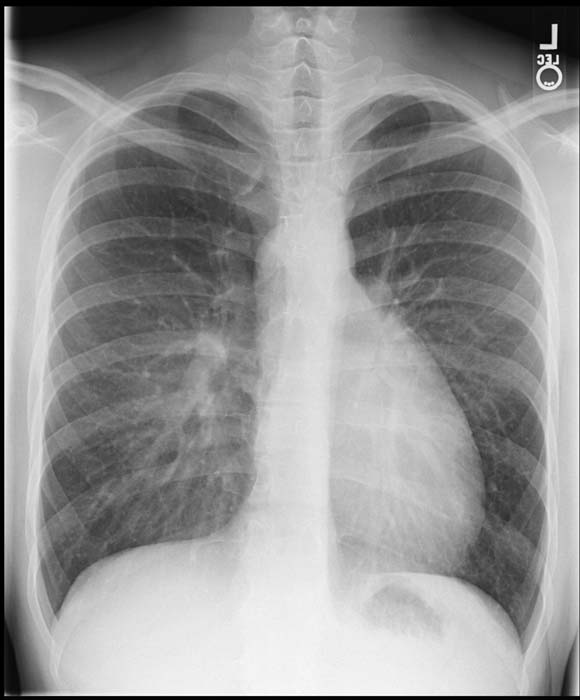
Overview
Chest Xray may show the classical egg-on-side pattern
Chest X Ray
- Generally, the superior mediastinum may be narrow due to the anterior-posterior relationship of the great vessels.
- Initially, cardiac size is normal, but soon enlarges with the cardiac apex shifted to the left and inferiorly, producing the typically ovale-shaped or egg-on-side pattern.
- If a VSD is present, there will be an increase of the pulmonar vascular margins.
-
Transposition of great vessels.
-

Transposition of great vessels.

