Dysfunctional uterine bleeding MRI: Difference between revisions
Jump to navigation
Jump to search
(Created page with " __NOTOC__ {{Dysfunctional uterine bleeding}} {{CMG}} {{AE}} {{VVS}} ==Overview== ==References== {{reflist|2}} {{WH}} {{WS}} Category:Needs content [[Category:Primary care...") |
m (→MRI) |
||
| (12 intermediate revisions by 3 users not shown) | |||
| Line 1: | Line 1: | ||
__NOTOC__ | |||
{{Dysfunctional uterine bleeding}} | {{Dysfunctional uterine bleeding}} | ||
{{CMG}} {{AE}} | {{CMG}}; {{AE}}[[User:AroojNaz|Arooj Naz, M.B.B.S]] | ||
==Overview== | ==Overview== | ||
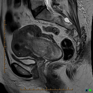
[[MRI]] is not commonly performed but it is considered the modality of choice for [[adenomyosis]]. Findings for other conditions may also be seen but may not be as reliable as other imaging studies. MRI can assist in furthering diagnosis [[metastasis]]. | |||
==MRI== | |||
{| class="wikitable" | |||
|+ | |||
MRI Findings | |||
!Underlying Cause | |||
!MRI | |||
!Findings | |||
|- | |||
|'''[[Endometrial polyp|Endometrial Polyps]]'''<ref name="“Radiopaedia”">{{cite web|url=https://radiopaedia.org/articles/endometrial-polyp}}</ref> | |||
|[[File:Prolapsed-endometrial-polyp-1.jpg|center|thumb|300x300px|Case courtesy of Dr Ammar Haouimi, Radiopaedia.org, rID: 70016]] | |||
| | |||
*[[Hypointense]] [[intracavitary]] masses | |||
*Surrounded by [[fluid]] and [[endometrium]] | |||
|- | |||
|'''[[Adenomyosis]]'''<ref name="“Radiopaedia”2">{{cite web|url=https://radiopaedia.org/articles/adenomyosis}}</ref> | |||
|[[File:Adenomyosis-with-endometrioma-1.jpg|center|thumb|333x333px|Case courtesy of Dr Varun Babu, Radiopaedia.org, rID: 43504]]<br /> | |||
|<br /> | |||
*Diagnosis of choice; [[Sensitivity (tests)|sensitivity]] of 78-88% and [[Specificity (tests)|specificity]] of 67-93% | |||
*Thickening of the [[junctional zone]] ≥12 mm (normal is up to 5 mm) | |||
*Appears as an ill-defined region of thickening | |||
|- | |||
|'''[[Leiomyoma]]'''<ref name="“Radiopaedia”3">{{cite web|url=https://radiopaedia.org/articles/uterine-leiomyoma}}</ref> | |||
| | |||
<br />[[File:Prolapsing-uterine-leiomyoma.jpg|center|thumb|300x300px|Case courtesy of Dr Chris O'Donnell, Radiopaedia.org, rID: 22883]]<br /> | |||
| | |||
*Only used for complex cases | |||
*May accurately determine size and location | |||
*Appear [[hypervascular]] | |||
*Helpful when surgery is considered or when trying to differentiate from fibroids | |||
|- | |||
|'''[[Uterine cancer|Malignancy]]'''<ref name="“Radiopaedia”4">{{cite web|url=https://radiopaedia.org/articles/endometrial-carcinoma}}</ref> | |||
| | |||
[[File:Endometrioid-adenocarcinoma-of-the-endometrium-2.jpg|center|thumb|300x300px|Case courtesy of Dr Bahman Rasuli, Radiopaedia.org, rID: 83200]] | |||
| | |||
*MRI may be helpful in improving accuracy for determining [[metastasis]] | |||
*Normal tissue enhances more than [[malignant tissue]] | |||
|- | |||
|'''[[PCOS]]'''<ref name="“Radiopaedia”5">{{cite web|url=https://radiopaedia.org/articles/polycystic-ovarian-syndrome-1}}</ref> | |||
|[[File:Polycystic-ovarian-syndrome-1.jpg|center|thumb|300x300px|Case courtesy of Dr Mostafa El-Feky, Radiopaedia.org, rID: 53010]] | |||
| | |||
*Not a routine examination for [[PCOS]] | |||
*F[[follicles]] will be seen on MRI | |||
|- | |||
|'''Endometrial Causes''' <ref name="“Radiopaedia”6">{{cite web|url=https://radiopaedia.org/articles/endometrioma1}}</ref> | |||
|[[File:Endometriosis-3.jpg|center|thumb|300x300px|Case courtesy of The Radswiki, Radiopaedia.org, rID: 11397]] | |||
| | |||
*[[Endometrioma]] appears hyperintense; the presence of hemorrhage results in a hypointense image | |||
|} | |||
==References== | ==References== | ||
{{reflist|2}} | {{reflist|2}} | ||
{{WH}} | {{WH}} | ||
{{WS}} | {{WS}} | ||
[[Category:Needs content]] | [[Category:Needs content]] | ||
[[Category:Disease]] | [[Category:Disease]] | ||
[[Category:Gynecology]] | [[Category:Gynecology]] | ||
Latest revision as of 01:22, 7 August 2022
|
Dysfunctional uterine bleeding Microchapters |
|
Differentiating Dysfunctional uterine bleeding from other Diseases |
|---|
|
Diagnosis |
|
Treatment |
|
Case Studies |
|
Dysfunctional uterine bleeding MRI On the Web |
|
American Roentgen Ray Society Images of Dysfunctional uterine bleeding MRI |
|
Directions to Hospitals Treating Dysfunctional uterine bleeding |
|
Risk calculators and risk factors for Dysfunctional uterine bleeding MRI |
Editor-In-Chief: C. Michael Gibson, M.S., M.D. [1]; Associate Editor(s)-in-Chief: Arooj Naz, M.B.B.S
Overview
MRI is not commonly performed but it is considered the modality of choice for adenomyosis. Findings for other conditions may also be seen but may not be as reliable as other imaging studies. MRI can assist in furthering diagnosis metastasis.
MRI
| Underlying Cause | MRI | Findings |
|---|---|---|
| Endometrial Polyps[1] |  |
|
| Adenomyosis[2] |  |
|
| Leiomyoma[3] |
 |
|
| Malignancy[4] |
 |
|
| PCOS[5] |  |
|
| Endometrial Causes [6] |  |
|
References
- ↑ https://radiopaedia.org/articles/endometrial-polyp. Missing or empty
|title=(help) - ↑ https://radiopaedia.org/articles/adenomyosis. Missing or empty
|title=(help) - ↑ https://radiopaedia.org/articles/uterine-leiomyoma. Missing or empty
|title=(help) - ↑ https://radiopaedia.org/articles/endometrial-carcinoma. Missing or empty
|title=(help) - ↑ https://radiopaedia.org/articles/polycystic-ovarian-syndrome-1. Missing or empty
|title=(help) - ↑ https://radiopaedia.org/articles/endometrioma1. Missing or empty
|title=(help)