|
|
| (133 intermediate revisions by 3 users not shown) |
| Line 1: |
Line 1: |
| | ==Physical examination== |
| | ==References== |
| | {{reflist|2}} |
| | |
| | {{WH}} |
| | {{WS}} |
| | |
| | ==References== |
| | {{Reflist|2}} |
| | |
| | |
| ===Pathophysiology prev=== | | ===Pathophysiology prev=== |
| <div style="-webkit-user-select: none;"> | | <div style="-webkit-user-select: none;"> |
| Line 10: |
Line 21: |
| {{CMG}} {{AE}} | | {{CMG}} {{AE}} |
|
| |
|
| ==Overview==
| |
|
| |
|
| {| class="wikitable"
| | ===Pathophysiology prev=== |
| ! rowspan="2" |Disease
| | <div style="-webkit-user-select: none;"> |
| ! colspan="7" |Symptoms
| | {| class="infobox" style="position: fixed; top: 65%; right: 10px; margin: 0 0 0 0; border: 0; float: right;" |
| ! rowspan="2" |Other features
| |
| ! colspan="2" |Diagnosis
| |
| |- | | |- |
| !Abdominal pain
| | | {{#ev:youtube|https://https://www.youtube.com/watch?v=5szNmKtyBW4|350}} |
| !Rectal pain
| |
| !Weightloss
| |
| !Fever
| |
| !Type of GI bleeding
| |
| !Diarrhea
| |
| !Constipation
| |
| !Laboratory findings
| |
| !Radio-Imaging findings
| |
| |- | |
| |[[Diverticulosis|'''Diverticulosis''']] | |
| | -
| |
| | -
| |
| | -
| |
| | -
| |
| |Red or maroon-colored [[blood]]
| |
| | -
| |
| | +
| |
| |
| |
| * Self limiting
| |
| | |
| * Seen in elderly
| |
| |Normal
| |
| |
| |
| Globular outpouchings on [[CT scan]]
| |
| |-
| |
| |[[Angiodysplasia|'''Angiodysplasia''']]
| |
| | -
| |
| | -
| |
| | -
| |
| | -
| |
| |Frank [[blood]]
| |
| | -
| |
| | -
| |
| |
| |
| * Painless [[bleeding]]
| |
| | |
| * [[Iron deficiency anemia]]
| |
| |Normal
| |
| |Normal
| |
| |-
| |
| |[[Hemorrhoids|'''Hemorrhoids''']]
| |
| | -
| |
| | +
| |
| | -
| |
| | -
| |
| |[[Blood]] on [[tissues]]
| |
| | -
| |
| | +
| |
| |
| |
| * [[Pain]] during [[defecation]]
| |
| | |
| * [[Anemia]]
| |
| | -
| |
| |Tortuous dilated vessels on [[anoscopy]]
| |
| |-
| |
| |[[Anal fissures|'''Anal fissures''']]
| |
| | -
| |
| | +
| |
| | -
| |
| | -
| |
| |[[Blood]] on [[tissues]]
| |
| | -
| |
| | +
| |
| |
| |
| * [[Pain]] during [[defecation]]
| |
| * [[Pain]] recurs with every [[bowel movement]]
| |
| |Normal except mild [[leucocytosis]]
| |
| |[[Anoscopy]]
| |
| |-
| |
| |[[Mesenteric Ischemia|'''Mesenteric Ischemia''']]
| |
| | +
| |
| | -
| |
| | +
| |
| | +
| |
| |Frank blood
| |
| | +
| |
| | -
| |
| |
| |
| * [[Pain]] alters with eating habits
| |
| | |
| * Associated with other comorbid conditions
| |
| |
| |
| * [[Leukocytosis]]
| |
| * Increased [[hematocrit]]
| |
| * [[High anion gap metabolic acidosis critical pathways|High anion gap metabolic acidosis]]
| |
| * [[Lactic acidosis]]
| |
| * [[Hyperphosphatemia|High phosphate levels]]
| |
| |
| |
| * [[Mesenteric]] [[edema]]
| |
| * [[Bowel]] dilatation
| |
| * Bowel wall thickening
| |
| * Intramural gas
| |
| * [[Mesenteric]] stranding
| |
| |-
| |
| |[[Ischemic colitis|'''Ischemic colitis''']]
| |
| | +
| |
| | -
| |
| | -
| |
| | +
| |
| |Frank blood
| |
| | +
| |
| | -
| |
| |3 phases
| |
| * [[Hyperactive]] phase
| |
| | |
| * [[Paralytic]] phase (absent bowel sounds)
| |
| | |
| * [[Shock]] phase
| |
| |
| |
| * [[Elevated white blood cell count]] more than 15,000/mm<sup>3</sup> in 20 patients (27%)
| |
| * The [[serum bicarbonate]] level was less than 24 mmol/L in 26 patients (36%)
| |
| |
| |
| * Mild moderate diffuse bowel wall thickening
| |
| * Marked hyperenhancement of the [[mucosa]]
| |
| |-
| |
| |[[Crohn's disease|'''Crohn's disease''']]
| |
| | +
| |
| | -
| |
| | +
| |
| | +
| |
| |Blood mixed with stools
| |
| | +
| |
| | +
| |
| |Extra intestinal manifestations
| |
| *[[Uveitis]]
| |
| * [[Sacroiliitis]]
| |
| * [[Anemia]]
| |
| * [[Peripheral neuropathy]]
| |
| |
| |
| * [[Anemia]]
| |
| * [[Leukocytosis]]
| |
| * [[Thrombocytosis]]
| |
| * [[Anti saccharomyces cerevisiae antibodies|Anti-Saccharomyces cerevisiae antibodies]]
| |
| |
| |
| * Skip lesions
| |
| * Bowel wall thickening
| |
| * Surrounding [[inflammation]], [[abscess]], and [[fistulae]]
| |
| |-
| |
| |[[Ulcerative colitis|'''Ulcerative colitis''']]
| |
| | +
| |
| | +
| |
| | +
| |
| | +
| |
| |Blood mixed with stools
| |
| | +
| |
| | +
| |
| |
| |
| * [[Joint swelling]]
| |
| * [[Joint pain]]
| |
| * [[Inflammation]] of the eye
| |
| * [[Skin]] involvement
| |
| * [[Fatty liver]]
| |
| * [[Thromboembolism]]
| |
| * [[Parenchymal lung disease]]
| |
| |
| |
| * [[Anemia]]
| |
| * [[Thrombocytosis]]
| |
| * A high [[platelet]] count
| |
| |
| |
| * Loss of the vascular appearance of the [[colon]]
| |
| * [[Erythema]] (or redness of the [[mucosa]]) and friability of the [[mucosa]]
| |
| * Superficial [[ulceration]], which may be confluent
| |
| * [[Polyp (medicine)|Pseudopolyps]]
| |
| |- | | |- |
| |[[Colon carcinoma|'''Colon carcinoma''']]
| |
| | +
| |
| | -†
| |
| | +
| |
| | +
| |
| |[[Fecal occult blood|Occult bleeding]]
| |
| | +
| |
| | +†
| |
| |
| |
| | + [[FOBT]] (fecal occult blood test)
| |
| ↑ [[CEA]]( and CA 19-9
| |
|
| |
| [[Hypercalcemia]]
| |
| |
| |
| * [[Biopsy]]
| |
| |} | | |} |
| | __NOTOC__ |
| | {{Cirrhosis}} |
| | {{CMG}} {{AE}} |
|
| |
|
| The following table differentiates all the diseases presenting with abdominal pain and lower gastrointestinal bleeding.
| | == History and Symptoms == |
|
| |
|
| <span style="font-size:85%">'''Abbreviations:'''
| | * History should include: |
| '''[[RUQ]]'''= Right upper quadrant of the abdomen, '''LUQ'''= Left upper quadrant, '''LLQ'''= Left lower quadrant, '''RLQ'''= Right lower quadrant, '''LFT'''= Liver function test, SIRS= [[Systemic inflammatory response syndrome]], '''[[ERCP]]'''= [[Endoscopic retrograde cholangiopancreatography]], '''IV'''= Intravenous, '''N'''= Normal, '''AMA'''= Anti mitochondrial antibodies, '''[[LDH]]'''= [[Lactate dehydrogenase]], '''GI'''= Gastrointestinal, '''CXR'''= Chest X ray, '''IgA'''= [[Immunoglobulin A]], '''IgG'''= [[Immunoglobulin G]], '''IgM'''= [[Immunoglobulin M]], '''CT'''= [[Computed tomography]], '''[[PMN]]'''= Polymorphonuclear cells, '''[[ESR]]'''= [[Erythrocyte sedimentation rate]], '''[[CRP]]'''= [[C-reactive protein]], TS= [[Transferrin saturation]], SF= Serum [[Ferritin]], SMA= [[Superior mesenteric artery]], SMV= [[Superior mesenteric vein]], ECG= [[Electrocardiogram]]</span>
| | ** Appearance of bowel movements |
| | ** Travel history |
| | ** Associated symptoms |
| | ** Immune status |
| | ** Woodland exposure |
| | ==References== |
| | {{reflist|2}} |
|
| |
|
| {| align="center" | | {{WH}} |
| |-
| | {{WS}} |
| |
| |
| {| style="border: 0px; font-size: 90%; margin: 3px;" align="center" | |
| ! rowspan="3" style="background:#4479BA; color: #FFFFFF;" align="center" |Disease
| |
| | colspan="13" rowspan="1" style="background:#4479BA; color: #FFFFFF;" align="center" |'''Clinical manifestations'''
| |
| ! colspan="2" rowspan="2" style="background:#4479BA; color: #FFFFFF;" align="center" |Diagnosis
| |
| ! rowspan="3" style="background:#4479BA; color: #FFFFFF;" align="center" |Comments
| |
| |-
| |
| | colspan="9" rowspan="1" style="background:#4479BA; color: #FFFFFF;" align="center" |'''Symptoms'''
| |
| ! colspan="4" rowspan="1" style="background:#4479BA; color: #FFFFFF;" align="center" | Signs
| |
| |-
| |
| ! style="background:#4479BA; color: #FFFFFF;" align="center" |Abdominal Pain
| |
| ! colspan="1" rowspan="1" style="background:#4479BA; color: #FFFFFF;" align="center" | Fever
| |
| ! style="background:#4479BA; color: #FFFFFF;" align="center" |Rigors and chills
| |
| ! style="background:#4479BA; color: #FFFFFF;" align="center" |Nausea or vomiting
| |
| ! style="background:#4479BA; color: #FFFFFF;" align="center" |Jaundice
| |
| ! style="background:#4479BA; color: #FFFFFF;" align="center" |Constipation
| |
| ! style="background:#4479BA; color: #FFFFFF;" align="center" |Diarrhea
| |
| ! style="background:#4479BA; color: #FFFFFF;" align="center" |Weight loss
| |
| ! style="background:#4479BA; color: #FFFFFF;" align="center" |GI bleeding
| |
| ! style="background:#4479BA; color: #FFFFFF;" align="center" |Hypo-
| |
| tension
| |
| ! colspan="1" rowspan="1" style="background:#4479BA; color: #FFFFFF;" align="center" | Guarding
| |
| ! style="background:#4479BA; color: #FFFFFF;" align="center" |Rebound Tenderness
| |
| ! style="background:#4479BA; color: #FFFFFF;" align="center" |Bowel sounds
| |
| ! colspan="1" rowspan="1" style="background:#4479BA; color: #FFFFFF;" align="center" | Lab Findings
| |
| ! style="background:#4479BA; color: #FFFFFF;" align="center" |Imaging
| |
| |-
| |
| | colspan="1" rowspan="1" style="padding: 5px 5px; background: #DCDCDC;" align="center" |[[Diverticulitis|Acute diverticulitis]]
| |
| | style="padding: 5px 5px; background: #F5F5F5;" align="center" |LLQ
| |
| | style="padding: 5px 5px; background: #F5F5F5;" align="center" | +
| |
| | style="padding: 5px 5px; background: #F5F5F5;" align="center" | ±
| |
| | style="padding: 5px 5px; background: #F5F5F5;" align="center" |<nowiki>+</nowiki>
| |
| | style="padding: 5px 5px; background: #F5F5F5;" align="center" | −
| |
| | style="padding: 5px 5px; background: #F5F5F5;" align="center" |<nowiki>+</nowiki>
| |
| | style="padding: 5px 5px; background: #F5F5F5;" align="center" | ±
| |
| | style="padding: 5px 5px; background: #F5F5F5;" align="center" |−
| |
| | style="padding: 5px 5px; background: #F5F5F5;" align="center" | +
| |
| | style="padding: 5px 5px; background: #F5F5F5;" align="center" | Positive in perforated diverticulitis
| |
| | style="padding: 5px 5px; background: #F5F5F5;" align="center" | +
| |
| | style="padding: 5px 5px; background: #F5F5F5;" align="center" | +
| |
| | style="padding: 5px 5px; background: #F5F5F5;" align="left" |Hypoactive
| |
| | style="padding: 5px 5px; background: #F5F5F5;" align="left" |
| |
| * [[Leukocytosis]]
| |
| | style="padding: 5px 5px; background: #F5F5F5;" align="left" |
| |
| * CT scan
| |
| * Ultrasound
| |
| | style="padding: 5px 5px; background: #F5F5F5;" align="left" |
| |
| * History of [[constipation]]
| |
| |-
| |
| | style="padding: 5px 5px; background: #DCDCDC;" align="center" |[[Inflammatory bowel disease]]
| |
| | style="padding: 5px 5px; background: #F5F5F5;" align="center" |Diffuse
| |
| | style="padding: 5px 5px; background: #F5F5F5;" align="center" | ±
| |
| | style="padding: 5px 5px; background: #F5F5F5;" align="center" | −
| |
| | style="padding: 5px 5px; background: #F5F5F5;" align="center" | −
| |
| | style="padding: 5px 5px; background: #F5F5F5;" align="center" | ±
| |
| | style="padding: 5px 5px; background: #F5F5F5;" align="center" | −
| |
| | style="padding: 5px 5px; background: #F5F5F5;" align="center" | +
| |
| | style="padding: 5px 5px; background: #F5F5F5;" align="center" |<nowiki>+</nowiki>
| |
| | style="padding: 5px 5px; background: #F5F5F5;" align="center" | +
| |
| | style="padding: 5px 5px; background: #F5F5F5;" align="center" | −
| |
| | style="padding: 5px 5px; background: #F5F5F5;" align="center" | −
| |
| | style="padding: 5px 5px; background: #F5F5F5;" align="center" | −
| |
| | style="padding: 5px 5px; background: #F5F5F5;" align="left" |Normal or hyperactive
| |
| | style="padding: 5px 5px; background: #F5F5F5;" align="left" |
| |
| * [[Anti-neutrophil cytoplasmic antibody]] ([[P-ANCA]]) in [[Ulcerative colitis]]
| |
| * [[Anti saccharomyces cerevisiae antibodies]] (ASCA) in [[Crohn's disease]]
| |
| | style="padding: 5px 5px; background: #F5F5F5;" align="left" |
| |
| * [[String sign]] on [[abdominal x-ray]] in [[Crohn's disease]]
| |
| | style="padding: 5px 5px; background: #F5F5F5;" align="left" |
| |
| Extra intestinal findings:
| |
| * [[Uveitis]]
| |
| * [[Arthritis]]
| |
| |-
| |
| | style="padding: 5px 5px; background: #DCDCDC;" align="center" |[[Infective colitis]]
| |
| | style="padding: 5px 5px; background: #F5F5F5;" align="center" |Diffuse
| |
| | style="padding: 5px 5px; background: #F5F5F5;" align="center" | +
| |
| | style="padding: 5px 5px; background: #F5F5F5;" align="center" | −
| |
| | style="padding: 5px 5px; background: #F5F5F5;" align="center" | ±
| |
| | style="padding: 5px 5px; background: #F5F5F5;" align="center" | −
| |
| | style="padding: 5px 5px; background: #F5F5F5;" align="center" | −
| |
| | style="padding: 5px 5px; background: #F5F5F5;" align="center" | +
| |
| | style="padding: 5px 5px; background: #F5F5F5;" align="center" | −
| |
| | style="padding: 5px 5px; background: #F5F5F5;" align="center" | +
| |
| | style="padding: 5px 5px; background: #F5F5F5;" align="center" | Positive in fulminant colitis
| |
| | style="padding: 5px 5px; background: #F5F5F5;" align="center" | ±
| |
| | style="padding: 5px 5px; background: #F5F5F5;" align="center" | ±
| |
| | style="padding: 5px 5px; background: #F5F5F5;" align="left" |Hyperactive
| |
| | style="padding: 5px 5px; background: #F5F5F5;" align="left" |
| |
| * [[Stool culture]] and studies
| |
| * Shiga toxin in bloody diarrhea
| |
| * [[PCR]]
| |
| | style="padding: 5px 5px; background: #F5F5F5;" align="left" |CT scan
| |
| * Bowel wall thickening
| |
| * Edema
| |
| | style="padding: 5px 5px; background: #F5F5F5;" align="left" |
| |
| |-
| |
| | style="padding: 5px 5px; background: #DCDCDC;" align="center" |Colon carcinoma
| |
| | style="padding: 5px 5px; background: #F5F5F5;" align="center" |Diffuse/localized
| |
| | style="padding: 5px 5px; background: #F5F5F5;" align="center" | −
| |
| | style="padding: 5px 5px; background: #F5F5F5;" align="center" | −
| |
| | style="padding: 5px 5px; background: #F5F5F5;" align="center" | −
| |
| | style="padding: 5px 5px; background: #F5F5F5;" align="center" | −
| |
| | style="padding: 5px 5px; background: #F5F5F5;" align="center" | ±
| |
| | style="padding: 5px 5px; background: #F5F5F5;" align="center" | ±
| |
| | style="padding: 5px 5px; background: #F5F5F5;" align="center" |<nowiki>+</nowiki>
| |
| | style="padding: 5px 5px; background: #F5F5F5;" align="center" | +
| |
| | style="padding: 5px 5px; background: #F5F5F5;" align="center" | ±
| |
| | style="padding: 5px 5px; background: #F5F5F5;" align="center" | −
| |
| | style="padding: 5px 5px; background: #F5F5F5;" align="center" | −
| |
| | style="padding: 5px 5px; background: #F5F5F5;" align="left" |
| |
| * Normal or hyperactive if obstruction present
| |
| | style="padding: 5px 5px; background: #F5F5F5;" align="left" |
| |
| * CBC
| |
| * Carcinoembryonic antigen (CEA)
| |
| | style="padding: 5px 5px; background: #F5F5F5;" align="left" |
| |
| * Colonoscopy
| |
| * Flexible sigmoidoscopy
| |
| * Barium enema
| |
| * CT colonography
| |
| | style="padding: 5px 5px; background: #F5F5F5;" align="left" |
| |
| * PILLCAM 2: A colon capsule for CRC screening may be used in patients with an incomplete colonoscopy who lacks obstruction
| |
| |-
| |
| | style="padding: 5px 5px; background: #DCDCDC;" align="center" |[[Hemochromatosis]]
| |
| | style="padding: 5px 5px; background: #F5F5F5;" align="center" |RUQ
| |
| | style="padding: 5px 5px; background: #F5F5F5;" align="center" | −
| |
| | style="padding: 5px 5px; background: #F5F5F5;" align="center" | −
| |
| | style="padding: 5px 5px; background: #F5F5F5;" align="center" | −
| |
| | style="padding: 5px 5px; background: #F5F5F5;" align="center" | −
| |
| | style="padding: 5px 5px; background: #F5F5F5;" align="center" | −
| |
| | style="padding: 5px 5px; background: #F5F5F5;" align="center" | −
| |
| | style="padding: 5px 5px; background: #F5F5F5;" align="center" | −
| |
| | style="padding: 5px 5px; background: #F5F5F5;" align="center" | Positive in cirrhotic patients
| |
| | style="padding: 5px 5px; background: #F5F5F5;" align="center" | −
| |
| | style="padding: 5px 5px; background: #F5F5F5;" align="center" | −
| |
| | style="padding: 5px 5px; background: #F5F5F5;" align="center" | −
| |
| | style="padding: 5px 5px; background: #F5F5F5;" align="left" |N
| |
| | style="padding: 5px 5px; background: #F5F5F5;" align="left" |
| |
| * >60% TS
| |
| * >240 μg/L SF
| |
| * Raised LFT <br>Hyperglycemia
| |
| | style="padding: 5px 5px; background: #F5F5F5;" align="left" |
| |
| * Ultrasound shows evidence of cirrhosis
| |
| | style="padding: 5px 5px; background: #F5F5F5;" align="left" |Extra intestinal findings:
| |
| * [[Hyperpigmentation]]
| |
| * [[Diabetes mellitus]]
| |
| * [[Arthralgia]]
| |
| * [[Erectile dysfunction|Impotence]] in males
| |
| * [[Cardiomyopathy]]
| |
| * [[Atherosclerosis]]
| |
| * [[Hypopituitarism]]
| |
| * [[Hypothyroidism]]
| |
| * Extrahepatic cancer
| |
| * Prone to specific infections
| |
| |-
| |
| | style="padding: 5px 5px; background: #DCDCDC;" align="center" |[[Mesenteric ischemia]]
| |
| | style="padding: 5px 5px; background: #F5F5F5;" align="center" |Periumbilical
| |
| | style="padding: 5px 5px; background: #F5F5F5;" align="center" |Positive if bowel becomes gangrenous
| |
| | style="padding: 5px 5px; background: #F5F5F5;" align="center" | −
| |
| | style="padding: 5px 5px; background: #F5F5F5;" align="center" |<nowiki>+</nowiki>
| |
| | style="padding: 5px 5px; background: #F5F5F5;" align="center" | −
| |
| | style="padding: 5px 5px; background: #F5F5F5;" align="center" | −
| |
| | style="padding: 5px 5px; background: #F5F5F5;" align="center" | +
| |
| | style="padding: 5px 5px; background: #F5F5F5;" align="center" |<nowiki>+</nowiki>
| |
| | style="padding: 5px 5px; background: #F5F5F5;" align="center" | +
| |
| | style="padding: 5px 5px; background: #F5F5F5;" align="center" | Positive if bowel becomes gangrenous
| |
| | style="padding: 5px 5px; background: #F5F5F5;" align="center" | Positive if bowel becomes gangrenous
| |
| | style="padding: 5px 5px; background: #F5F5F5;" align="center" | −
| |
| | style="padding: 5px 5px; background: #F5F5F5;" align="left" |Hyperactive to absent
| |
| | style="padding: 5px 5px; background: #F5F5F5;" align="left" |
| |
| * [[Leukocytosis]] and [[lactic acidosis]]
| |
| * [[Amylase]] levels
| |
| * [[D-dimer]]
| |
| | style="padding: 5px 5px; background: #F5F5F5;" align="left" |CT angiography
| |
| * SMA or SMV thrombosis
| |
| | style="padding: 5px 5px; background: #F5F5F5;" align="left" |
| |
| * Also known as abdominal angina that worsens with eating
| |
| |-
| |
| | style="padding: 5px 5px; background: #DCDCDC;" align="center" |[[Ischemic colitis|Acute ischemic colitis]]
| |
| | style="padding: 5px 5px; background: #F5F5F5;" align="center" | Diffuse
| |
| | style="padding: 5px 5px; background: #F5F5F5;" align="center" | +
| |
| | style="padding: 5px 5px; background: #F5F5F5;" align="center" | ±
| |
| | style="padding: 5px 5px; background: #F5F5F5;" align="center" |<nowiki>+</nowiki>
| |
| | style="padding: 5px 5px; background: #F5F5F5;" align="center" | −
| |
| | style="padding: 5px 5px; background: #F5F5F5;" align="center" | −
| |
| | style="padding: 5px 5px; background: #F5F5F5;" align="center" | +
| |
| | style="padding: 5px 5px; background: #F5F5F5;" align="center" |<nowiki>+</nowiki>
| |
| | style="padding: 5px 5px; background: #F5F5F5;" align="center" | +
| |
| | style="padding: 5px 5px; background: #F5F5F5;" align="center" | +
| |
| | style="padding: 5px 5px; background: #F5F5F5;" align="center" |<nowiki>+</nowiki>
| |
| | style="padding: 5px 5px; background: #F5F5F5;" align="center" |<nowiki>+</nowiki>
| |
| | style="padding: 5px 5px; background: #F5F5F5;" align="left" |Hyperactive then absent
| |
| | style="padding: 5px 5px; background: #F5F5F5;" align="left" |
| |
| * [[Leukocytosis]]
| |
| | style="padding: 5px 5px; background: #F5F5F5;" align="left" |[[Abdominal x-ray]]
| |
| * Distension and pneumatosis
| |
| CT scan
| |
| * Double halo appearance, thumbprinting
| |
| * Thickening of bowel
| |
| | style="padding: 5px 5px; background: #F5F5F5;" align="left" |
| |
| * May lead to shock
| |
| |-
| |
| | style="padding: 5px 5px; background: #DCDCDC;" align="center" |[[Ruptured abdominal aortic aneurysm]]
| |
| | style="padding: 5px 5px; background: #F5F5F5;" align="center" | Diffuse
| |
| | style="padding: 5px 5px; background: #F5F5F5;" align="center" | ±
| |
| | style="padding: 5px 5px; background: #F5F5F5;" align="center" | −
| |
| | style="padding: 5px 5px; background: #F5F5F5;" align="center" |<nowiki>+</nowiki>
| |
| | style="padding: 5px 5px; background: #F5F5F5;" align="center" | −
| |
| | style="padding: 5px 5px; background: #F5F5F5;" align="center" | −
| |
| | style="padding: 5px 5px; background: #F5F5F5;" align="center" | −
| |
| | style="padding: 5px 5px; background: #F5F5F5;" align="center" |<nowiki>+</nowiki>
| |
| | style="padding: 5px 5px; background: #F5F5F5;" align="center" | +
| |
| | style="padding: 5px 5px; background: #F5F5F5;" align="center" | +
| |
| | style="padding: 5px 5px; background: #F5F5F5;" align="center" | −
| |
| | style="padding: 5px 5px; background: #F5F5F5;" align="center" | −
| |
| | style="padding: 5px 5px; background: #F5F5F5;" align="left" |N
| |
| | style="padding: 5px 5px; background: #F5F5F5;" align="left" |
| |
| * [[Fibrinogen]]
| |
| * [[D-dimer]]
| |
| | style="padding: 5px 5px; background: #F5F5F5;" align="left" |
| |
| * Focused Assessment with Sonography in Trauma (FAST)
| |
| | style="padding: 5px 5px; background: #F5F5F5;" align="left" |
| |
| * Unstable hemodynamics
| |
| |-
| |
| | style="padding: 5px 5px; background: #DCDCDC;" align="center" |Intra-abdominal or [[retroperitoneal hemorrhage]]
| |
| | style="padding: 5px 5px; background: #F5F5F5;" align="center" | Diffuse
| |
| | style="padding: 5px 5px; background: #F5F5F5;" align="center" | ±
| |
| | style="padding: 5px 5px; background: #F5F5F5;" align="center" | −
| |
| | style="padding: 5px 5px; background: #F5F5F5;" align="center" | ±
| |
| | style="padding: 5px 5px; background: #F5F5F5;" align="center" | −
| |
| | style="padding: 5px 5px; background: #F5F5F5;" align="center" | −
| |
| | style="padding: 5px 5px; background: #F5F5F5;" align="center" | −
| |
| | style="padding: 5px 5px; background: #F5F5F5;" align="center" | −
| |
| | style="padding: 5px 5px; background: #F5F5F5;" align="center" | +
| |
| | style="padding: 5px 5px; background: #F5F5F5;" align="center" | +
| |
| | style="padding: 5px 5px; background: #F5F5F5;" align="center" | −
| |
| | style="padding: 5px 5px; background: #F5F5F5;" align="center" | −
| |
| | style="padding: 5px 5px; background: #F5F5F5;" align="left" |N
| |
| | style="padding: 5px 5px; background: #F5F5F5;" align="left" |
| |
| * ↓ Hb
| |
| * ↓ Hct
| |
| | style="padding: 5px 5px; background: #F5F5F5;" align="left" |
| |
| * CT scan
| |
| | style="padding: 5px 5px; background: #F5F5F5;" align="left" |
| |
| * History of [[trauma]]
| |
| |-
| |
| |}
| |
| |}
| |
| Cirrhosis occurs due to long term [[liver]] injury which causes an imbalance between [[matrix]] production and degradation. Early disruption of the normal hepatic matrix results in its replacement by [[scar tissue]], which in turn has deleterious effects on cell function.
| |
|
| |
|
| | ==Other Imaging Findings== |
| | * [[Endoscopy]] |
| | * [[Barium enema]] |
| | * [[Colonoscopy]] |
| | * [[Sigmoidoscopy]] |
|
| |
|
| Surgery
| | ==Other diagnostic studies== |
| Incidentally discovered Meckel diverticulum
| | == Other Diagnostic Studies == |
| When a Meckel diverticulum is incidentally discovered at laparoscopy or laparotomy, the surgeon must decide whether to resect. Most surgeons generally do not resect a diverticulum with a wide mouth. However, a diverticulum with a narrow neck, which may obstruct or twist, can be easily resected at the neck without the need for segmental resection. A diverticulum deemed abnormal because of inflammation, thickening, or intramural pathology should be resected, with the decision for local or segmental resection based on the pathology. [5]
| |
|
| |
|
| In 1976, Soltero and Bill reported that the lifetime risk of complications from Meckel diverticulum was 4.2% and that the risk decreased with age. [9] Their data indicated that 800 asymptomatic diverticula would have to be removed to save the life of one patient. On that basis, they opposed surgical excision.
| | * Breath hydrogen test |
|
| |
|
| Miltiadis et al stated that resection of incidentally found Meckel diverticulum protected patients from future surgery caused by complications of Meckel diverticulum. This strategy could help reduce morbidity and mortality, especially in elderly patients.
| | * [[HIV test]]ing for those patients suspected of having HIV |
|
| |
|
| Cullen et al reported that prophylactic diverticulectomy is warranted to eliminate the possibility of future deaths. [3] As a result, they advocated diverticulectomy for asymptomatic disease even in the older age group.
| | == |
|
| |
|
| Overall, selected cases of incidentally discovered diverticula in adults and most asymptomatic diverticula found in children [10] may be removed. Resection is recommended in the following cases:
| | ==Overview== |
| | |
| Patients younger than 40 years
| |
| Diverticula longer than 2 cm
| |
| Diverticula with narrow necks
| |
| Diverticula with fibrous bands
| |
| Suspected ectopic gastric tissue
| |
| Inflamed, thickened diverticula
| |
| Removal of a healthy diverticulum in the presence of peritonitis, Crohn disease, ulcerative colitis, or any other complication that would militate against resection is not advised.
| |
| | |
| | |
| | |
| ==Meckel== | |
| <gallery>
| |
| Image:
| |
| | |
| Positive-meckels-scan-001.jpg
| |
| | |
| </gallery>
| |
| <gallery>
| |
| Image:
| |
| | |
| Meckel's-diverticulitis-001.jpg
| |
| | |
| Image:
| |
| | |
| Meckel's-diverticulitis-002.jpg
| |
| | |
| Image:
| |
| | |
| Meckel's-diverticulitis-003.jpg
| |
| | |
| Image:
| |
| | |
| Meckel's-diverticulitis-004.jpg
| |
| | |
| </gallery>
| |
|
| |
|
| ==References== | | ==References== |
| {{reflist|2}} | | {{reflist|2}} |
|
| |
|
| [[Category:Gastroenterology]]
| | {{WH}} |
| [[Category:Hepatology]]
| |
| [[Category:Disease]]
| |
| | |
| {{WS}} | | {{WS}} |
| {{WH}}
| |
| ===the end===
| |
| -
| |
| ==== Portal HTN results from the combination of the following: ====
| |
| * Structural disturbances associated with advanced liver disease account for 70% of total hepatic vascular resistance.
| |
| * Functional abnormalities such as endothelial dysfunction and increased hepatic vascular tone account for 30% of total hepatic vascular resistance.
| |
| Pathogenesis of Cirrhosis due to Alcohol:
| |
| * More than 66 percent of all American adults consume alcohol.
| |
| * Cirrhosis due to alcohol accounts for approximately forty percent of mortality rates due to cirrhosis.
| |
| * Mechanisms of alcohol-induced damage include:
| |
| ** Impaired protein synthesis, secretion, glycosylation
| |
| * Ethanol intake leads to elevated accumulation of intracellular triglycerides by:
| |
| ** Lipoprotein secretion
| |
| ** Decreased fatty acid oxidation
| |
| ** Increased fatty acid uptake
| |
| * Alcohol is converted by Alcohol dehydrogenase to acetaldehyde.
| |
| * Due to the high reactivity of acetaldehyde, it forms acetaldehyde-protein adducts which cause damage to cells by:
| |
| ** Trafficking of hepatic proteins
| |
| ** Interrupting microtubule formation
| |
| ** Interfering with enzyme activities
| |
| * Damage of hepatocytes leads to the formation of reactive oxygen species that activate Kupffer cells.<ref name="pmid11984538">{{cite journal |vauthors=Arthur MJ |title=Reversibility of liver fibrosis and cirrhosis following treatment for hepatitis C |journal=Gastroenterology |volume=122 |issue=5 |pages=1525–8 |year=2002 |pmid=11984538 |doi= |url=}}</ref>
| |
| *Kupffer cell activation leads to the production of profibrogenic cytokines that stimulates stellate cells.
| |
| *Stellate cell activation leads to the production of extracellular matrix and collagen.
| |
| * Portal triads develop connections with central veins due to connective tissue formation in pericentral and periportal zones, leading to the formation of regenerative nodules.
| |
| * Shrinkage of the liver occurs over years due to repeated insults that lead to:
| |
| ** Loss of hepatocytes
| |
| ** Increased production and deposition of collagen
| |
|
| |
|
| | | ===Pathophysiology prev=== |
| Pathology
| | <div style="-webkit-user-select: none;"> |
| * There are four stages of Cirrhosis as it progresses:
| | {| class="infobox" style="position: fixed; top: 65%; right: 10px; margin: 0 0 0 0; border: 0; float: right;" |
| ** Chronic nonsuppurative destructive cholangitis - inflammation and necrosis of portal tracts with lymphocyte infiltration leading to the destruction of the bile ducts.
| |
| ** Development of biliary stasis and fibrosis
| |
| *Periportal fibrosis progresses to bridging fibrosis
| |
| *Increased proliferation of smaller bile ductules leading to regenerative nodule formation.
| |
| | |
| | |
| | |
| ===Causes=== | |
| {| class="wikitable"
| |
| ! style="background:#4479BA; color: #FFFFFF;" align="center" + |Drugs and Toxins
| |
| ! style="background:#4479BA; color: #FFFFFF;" align="center" + |Infections
| |
| ! style="background:#4479BA; color: #FFFFFF;" align="center" + |Autoimmune
| |
| ! style="background:#4479BA; color: #FFFFFF;" align="center" + |Metabolic
| |
| ! style="background:#4479BA; color: #FFFFFF;" align="center" + |Biliary obstruction(Secondary bilary cirrhosis)
| |
| ! style="background:#4479BA; color: #FFFFFF;" align="center" + |Vascular
| |
| ! style="background:#4479BA; color: #FFFFFF;" align="center" + |Miscellaneous
| |
| |- | | |- |
| |Alcohol | | | {{#ev:youtube|https://https://www.youtube.com/watch?v=5szNmKtyBW4|350}} |
| |Hepatitis B | |
| |Primary Biliary Cirrhosis | |
| |Wilson's disease
| |
| |Cystic fibrosis
| |
| |Chronic RHF
| |
| |Sarcoidosis
| |
| |- | | |- |
| |Methotrexate
| |
| |Hepatitis C
| |
| |Autoimmune hepatitis
| |
| |Hemochromatosis
| |
| |Biliary atresia
| |
| |Budd-Chiari syndrome
| |
| |Intestinal
| |
|
| |
| bypass operations for obesity
| |
| |-
| |
| |Isoniazid
| |
| |Schistosoma japonicum
| |
| |Primary Sclerosing Cholangitis
| |
| |Alpha-1 antitrypsin deficiency
| |
| |Bile duct strictures
| |
| |Veno-occlusive disease
| |
| |Cryptogenic: unknown
| |
| |-
| |
| |Methyldopa
| |
| |
| |
| |
| |
| |Porphyria
| |
| |Gallstones
| |
| |
| |
| |
| |
| |-
| |
| |
| |
| |
| |
| |
| |
| |Glycogen storage diseases (such as Galactosaemia, Abetalipoproteinaemia)
| |
| |
| |
| |
| |
| |
| |
| |} | | |} |
| | | __NOTOC__ |
| ===Cirrhosis===
| | {{Cirrhosis}} |
| | | {{CMG}} {{AE}} |
| Pathophysiology <ref name="pmid7932316">{{cite journal |vauthors=Arthur MJ, Iredale JP |title=Hepatic lipocytes, TIMP-1 and liver fibrosis |journal=J R Coll Physicians Lond |volume=28 |issue=3 |pages=200–8 |year=1994 |pmid=7932316 |doi= |url=}}</ref><ref name="pmid8502273">{{cite journal |vauthors=Friedman SL |title=Seminars in medicine of the Beth Israel Hospital, Boston. The cellular basis of hepatic fibrosis. Mechanisms and treatment strategies |journal=N. Engl. J. Med. |volume=328 |issue=25 |pages=1828–35 |year=1993 |pmid=8502273 |doi=10.1056/NEJM199306243282508 |url=}}</ref><ref name="pmid8682489">{{cite journal |vauthors=Iredale JP |title=Matrix turnover in fibrogenesis |journal=Hepatogastroenterology |volume=43 |issue=7 |pages=56–71 |year=1996 |pmid=8682489 |doi= |url=}}</ref><ref name="pmid7959178">{{cite journal |vauthors=Gressner AM |title=Perisinusoidal lipocytes and fibrogenesis |journal=Gut |volume=35 |issue=10 |pages=1331–3 |year=1994 |pmid=7959178 |pmc=1374996 |doi= |url=}}</ref><ref name="pmid17332881">{{cite journal |vauthors=Iredale JP |title=Models of liver fibrosis: exploring the dynamic nature of inflammation and repair in a solid organ |journal=J. Clin. Invest. |volume=117 |issue=3 |pages=539–48 |year=2007 |pmid=17332881 |pmc=1804370 |doi=10.1172/JCI30542 |url=}}</ref><ref name="pmid11984538">{{cite journal |vauthors=Arthur MJ |title=Reversibility of liver fibrosis and cirrhosis following treatment for hepatitis C |journal=Gastroenterology |volume=122 |issue=5 |pages=1525–8 |year=2002 |pmid=11984538 |doi= |url=}}</ref>
| |
| * When an injured issue is replaced by a collagenous scar, it is termed as fibrosis.
| |
| * When fibrosis of the liver reaches an advanced stage where distortion of the hepatic vasculature also occurs, it is termed as cirrhosis of the liver.
| |
| * The cellular mechanisms responsible for cirrhosis are similar regardless of the type of initial insult and site of injury within the liver lobule.
| |
| * Viral hepatitis involves the periportal region, whereas involvement in alcoholic liver disease is largely pericentral.
| |
| * If the damage progresses, panlobular cirrhosis may result.
| |
| * Cirrhosis involves the following steps: <ref name="pmid7737629">{{cite journal |vauthors=Wanless IR, Wong F, Blendis LM, Greig P, Heathcote EJ, Levy G |title=Hepatic and portal vein thrombosis in cirrhosis: possible role in development of parenchymal extinction and portal hypertension |journal=Hepatology |volume=21 |issue=5 |pages=1238–47 |year=1995 |pmid=7737629 |doi= |url=}}</ref>
| |
| ** Inflammation
| |
| ** Hepatic stellate cell activation
| |
| ** Angiogenesis
| |
| ** Fibrogenesis
| |
| * Kupffer cells are hepatic macrophages responsible for Hepatic Stellate cell activation during injury.
| |
| * The hepatic stellate cell (also known as the perisinusoidal cell or Ito cell) plays a key role in the pathogenesis of liver fibrosis/cirrhosis.
| |
| * Hepatic stellate cells(HSC) are usually located in the subendothelial space of Disse and become activated to a myofibroblast-like phenotype in areas of liver injury.
| |
| * Collagen and non collagenous matrix proteins responsible for fibrosis are produced by the activated Hepatic Stellate Cells(HSC).
| |
| * Hepatocyte damage causes the release of lipid peroxidases from injured cell membranes leading to necrosis of parenchymal cells.
| |
| * Activated HSC produce numerous cytokines and their receptors, such as PDGF and TGF-f31 which are responsible for fibrogenesis.
| |
| * The matrix formed due to HSC activation is deposited in the space of Disse and leads to loss of fenestrations of endothelial cells, which is a process called capillarization.
| |
| * Cirrhosis leads to hepatic microvascular changes characterised by <ref name="pmid19157625">{{cite journal |vauthors=Fernández M, Semela D, Bruix J, Colle I, Pinzani M, Bosch J |title=Angiogenesis in liver disease |journal=J. Hepatol. |volume=50 |issue=3 |pages=604–20 |year=2009 |pmid=19157625 |doi=10.1016/j.jhep.2008.12.011 |url=}}</ref>
| |
| ** formation of intra hepatic shunts (due to angiogenesis and loss of parenchymal cells)
| |
| ** hepatic endothelial dysfunction
| |
| * The endothelial dysfunction is characterised by <ref name="pmid22504334">{{cite journal |vauthors=García-Pagán JC, Gracia-Sancho J, Bosch J |title=Functional aspects on the pathophysiology of portal hypertension in cirrhosis |journal=J. Hepatol. |volume=57 |issue=2 |pages=458–61 |year=2012 |pmid=22504334 |doi=10.1016/j.jhep.2012.03.007 |url=}}</ref>
| |
| ** insufficient release of vasodilators, such as nitric oxide due to oxidative stress
| |
| ** increased production of vasoconstrictors (mainly adrenergic stimulation and activation of endothelins and RAAS)
| |
| * Fibrosis eventually leads to formation of septae that grossly distort the liver architecture which includes both the liver parenchyma and the vasculature. A cirrhotic liver compromises hepatic sinusoidal exchange by shunting arterial and portal blood directly into the central veins (hepatic outflow). Vascularized fibrous septa connect central veins with portal tracts leading to islands of hepatocytes surrounded by fibrous bands without central veins.<ref name="pmid18328931">{{cite journal |vauthors=Schuppan D, Afdhal NH |title=Liver cirrhosis |journal=Lancet |volume=371 |issue=9615 |pages=838–51 |year=2008 |pmid=18328931 |pmc=2271178 |doi=10.1016/S0140-6736(08)60383-9 |url=}}</ref><ref name="pmid15094237">{{cite journal |vauthors=Desmet VJ, Roskams T |title=Cirrhosis reversal: a duel between dogma and myth |journal=J. Hepatol. |volume=40 |issue=5 |pages=860–7 |year=2004 |pmid=15094237 |doi=10.1016/j.jhep.2004.03.007 |url=}}</ref><ref name="pmid11079009">{{cite journal |vauthors=Wanless IR, Nakashima E, Sherman M |title=Regression of human cirrhosis. Morphologic features and the genesis of incomplete septal cirrhosis |journal=Arch. Pathol. Lab. Med. |volume=124 |issue=11 |pages=1599–607 |year=2000 |pmid=11079009 |doi=10.1043/0003-9985(2000)124<1599:ROHC>2.0.CO;2 |url=}}</ref>
| |
| * The formation of fibrotic bands is accompanied by regenerative nodule formation in the hepatic parenchyma.
| |
| * Advancement of cirrhosis may lead to parenchymal dysfunction and development of portal hypertension.
| |
| * Portal HTN results from the combination of the following:
| |
| ** Structural disturbances associated with advanced liver disease account for 70% of total hepatic vascular resistance.
| |
| ** Functional abnormalities such as endothelial dysfunction and increased hepatic vascular tone account for 30% of total hepatic vascular resistance.
| |
| | |
| Pathogenesis of Cirrhosis due to Alcohol:
| |
| * More than 66 percent of all American adults consume alcohol.
| |
| * Cirrhosis due to alcohol accounts for approximately forty percent of mortality rates due to cirrhosis.
| |
| * Mechanisms of alcohol-induced damage include:
| |
| ** Impaired protein synthesis, secretion, glycosylation
| |
| * Ethanol intake leads to elevated accumulation of intracellular triglycerides by:
| |
| ** Lipoprotein secretion
| |
| ** Decreased fatty acid oxidation
| |
| ** Increased fatty acid uptake
| |
| * Alcohol is converted by Alcohol dehydrogenase to acetaldehyde.
| |
| * Due to the high reactivity of acetaldehyde, it forms acetaldehyde-protein adducts which cause damage to cells by:
| |
| ** Trafficking of hepatic proteins
| |
| ** Interrupting microtubule formation
| |
| ** Interfering with enzyme activities
| |
| * Damage of hepatocytes leads to the formation of reactive oxygen species that activate Kupffer cells.<ref name="pmid11984538">{{cite journal |vauthors=Arthur MJ |title=Reversibility of liver fibrosis and cirrhosis following treatment for hepatitis C |journal=Gastroenterology |volume=122 |issue=5 |pages=1525–8 |year=2002 |pmid=11984538 |doi= |url=}}</ref>
| |
| *Kupffer cell activation leads to the production of profibrogenic cytokines that stimulates stellate cells.
| |
| *Stellate cell activation leads to the production of extracellular matrix and collagen.
| |
| * Portal triads develop connections with central veins due to connective tissue formation in pericentral and periportal zones, leading to the formation of regenerative nodules.
| |
| * Shrinkage of the liver occurs over years due to repeated insults that lead to:
| |
| ** Loss of hepatocytes
| |
| ** Increased production and deposition of collagen
| |
| | |
| | |
| Pathology
| |
| * There are four stages of Cirrhosis as it progresses:
| |
| ** Chronic nonsuppurative destructive cholangitis - inflammation and necrosis of portal tracts with lymphocyte infiltration leading to the destruction of the bile ducts.
| |
| ** Development of biliary stasis and fibrosis
| |
| *Periportal fibrosis progresses to bridging fibrosis
| |
| *Increased proliferation of smaller bile ductules leading to regenerative nodule formation.
| |
|
| |
|
| ==Video codes== | | ==Video codes== |
| Line 671: |
Line 85: |
| ===Normal video=== | | ===Normal video=== |
| {{#ev:youtube|x6e9Pk6inYI}} | | {{#ev:youtube|x6e9Pk6inYI}} |
| | {{#ev:youtube|4uSSvD1BAHg}} |
| | {{#ev:youtube|PQXb5D-5UZw}} |
| | {{#ev:youtube|UVJYQlUm2A8}} |
|
| |
|
| ===Video in table=== | | ===Video in table=== |
| Line 702: |
Line 119: |
| ===Image and text to the right=== | | ===Image and text to the right=== |
|
| |
|
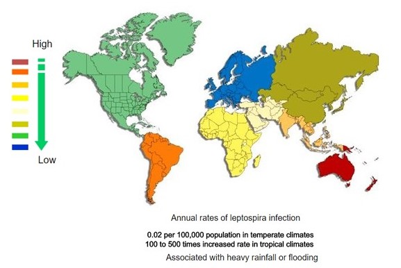
| <figure-inline><figure-inline><figure-inline><figure-inline><figure-inline><figure-inline><figure-inline><figure-inline><figure-inline><figure-inline><figure-inline><figure-inline><figure-inline><figure-inline><figure-inline>[[File:Global distribution of leptospirosis.jpg|577x577px]]</figure-inline></figure-inline></figure-inline></figure-inline></figure-inline></figure-inline></figure-inline></figure-inline></figure-inline></figure-inline></figure-inline></figure-inline></figure-inline></figure-inline></figure-inline> Recent out break of leptospirosis is reported in Bronx, New York and found 3 cases in the months January and February, 2017. | | <figure-inline><figure-inline><figure-inline><figure-inline><figure-inline><figure-inline><figure-inline><figure-inline><figure-inline><figure-inline><figure-inline><figure-inline><figure-inline><figure-inline><figure-inline><figure-inline><figure-inline><figure-inline><figure-inline><figure-inline><figure-inline><figure-inline><figure-inline><figure-inline><figure-inline><figure-inline><figure-inline>[[File:Global distribution of leptospirosis.jpg|577x577px]]</figure-inline></figure-inline></figure-inline></figure-inline></figure-inline></figure-inline></figure-inline></figure-inline></figure-inline></figure-inline></figure-inline></figure-inline></figure-inline></figure-inline></figure-inline></figure-inline></figure-inline></figure-inline></figure-inline></figure-inline></figure-inline></figure-inline></figure-inline></figure-inline></figure-inline></figure-inline></figure-inline> Recent out break of leptospirosis is reported in Bronx, New York and found 3 cases in the months January and February, 2017. |
|
| |
|
| ===Gallery=== | | ===Gallery=== |
| Line 717: |
Line 134: |
| ==References== | | ==References== |
| {{Reflist|2}} | | {{Reflist|2}} |
|
| |
| [[Category:Gastroenterology]]
| |
| [[Category:Hepatology]]
| |
| [[Category:Disease]]
| |
|
| |
| {{WS}} | | {{WS}} |
| {{WH}} | | {{WH}} |
| Line 728: |
Line 140: |
| REFERENCES | | REFERENCES |
| <references /> | | <references /> |
| | |
| | [[Category:Gastroenterology]] |
| | [[Category:Needs overview]] |
| | [[Category:Hepatology]] |
| | [[Category:Disease]] |


 </figure-inline></figure-inline></figure-inline></figure-inline></figure-inline></figure-inline></figure-inline></figure-inline></figure-inline></figure-inline></figure-inline></figure-inline></figure-inline></figure-inline></figure-inline></figure-inline></figure-inline></figure-inline></figure-inline></figure-inline></figure-inline></figure-inline></figure-inline></figure-inline></figure-inline></figure-inline></figure-inline> Recent out break of leptospirosis is reported in Bronx, New York and found 3 cases in the months January and February, 2017.
![Histopathology of a pancreatic endocrine tumor (insulinoma). Source:https://librepathology.org/wiki/Neuroendocrine_tumour_of_the_pancreas[1]](/images/2/2f/Pancreatic_insulinoma_histology_2.JPG)
![Histopathology of a pancreatic endocrine tumor (insulinoma). Chromogranin A immunostain. Source:https://librepathology.org/wiki/Neuroendocrine_tumour_of_the_pancreas[1]](/images/a/a3/Pancreatic_insulinoma_histopathology_3.JPG)
![Histopathology of a pancreatic endocrine tumor (insulinoma). Insulin immunostain. Source:https://librepathology.org/wiki/Neuroendocrine_tumour_of_the_pancreas[1]](/images/d/d5/Pancreatic_insulinoma_histology_4.JPG)