|
|
| (21 intermediate revisions by one other user not shown) |
| Line 1: |
Line 1: |
| | | ==pic== |
| {| | | {| |
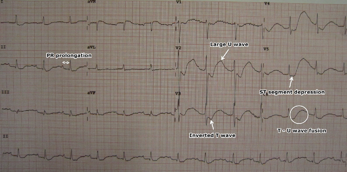
| |[[image:LowKECG.png|thumb|700px|center|An ECG in a person with a potassium level of 1.1 showing the classical ECG changes of ST segment depression, inverted T waves, large U waves, and a slightly prolonged PR interval. By James Heilman, MD - Own work, CC BY-SA 3.0<ref name="urlFile:LowKECG.JPG - Wikimedia Commons">{{cite web |url=https://commons.wikimedia.org/w/index.php?curid=12210926 |title=File:LowKECG.JPG - Wikimedia Commons |format= |work= |accessdate=}}</ref>]] | | |[[image:LowKECG.png|thumb|700px|center|An ECG in a person with a potassium level of 1.1 showing the classical ECG changes of ST segment depression, inverted T waves, large U waves, and a slightly prolonged PR interval. By James Heilman, MD - Own work, CC BY-SA 3.0]] |
| |} | | |} |
| <br style="clear:left" /> | | <br style="clear:left" /> |
|
| |
|
| [[image:LowKECG.png|thumb|700px|center|An ECG in a person with a potassium level of 1.1 showing the classical ECG changes of ST segment depression, inverted T waves, large U waves, and a slightly prolonged PR interval. By James Heilman, MD - Own work, CC BY-SA 3.0<ref name="urlFile:LowKECG.JPG - Wikimedia Commons">{{cite web |url=https://commons.wikimedia.org/w/index.php?curid=12210926 |title=File:LowKECG.JPG - Wikimedia Commons |format= |work= |accessdate=}}</ref>]] | | [[image:LowKECG.png|thumb|700px|right|An ECG in a person with a potassium level of 1.1 showing the classical ECG changes of ST segment depression, inverted T waves, large U waves, and a slightly prolonged PR interval. By James Heilman, MD - Own work, CC BY-SA 3.0]] |
| <br style="clear:left" /> | | <br style="clear:left" /> |
|
| |
|
| Line 19: |
Line 19: |
| | align="left" style="background:#F5F5F5;" + | | | | align="left" style="background:#F5F5F5;" + | |
| *[[Lung]] | | *[[Lung]] |
| *[[Kidney]]
| | | align="center" style="background:#F5F5F5;" + | |
| *[[Breast]]
| |
| *[[Urinary bladder|Bladder]]
| |
| *[[Endometrium|Endometrial]]
| |
| *[[Cervical]]
| |
| *[[Thyroid]]
| |
| *[[Lymphoma]]
| |
| *[[Multiple myeloma|Myeloma]]
| |
| *[[Brain]]
| |
| | align="left" style="background:#F5F5F5;" + | 5-7 fold more risks than the general population | |
| *[[Lung]] | | *[[Lung]] |
| *[[Ovary|Ovarian]]
| |
| *[[Breast]]
| |
| *[[Colon (anatomy)|Colorectal]]
| |
| *[[Cervical]]
| |
| *[[Urinary bladder|Bladder]]
| |
| *[[Nasopharynx|Nasopharyngeal]]
| |
| *[[Esophageal]]
| |
| *[[Pancreas|Pancreatic]]
| |
| *[[Kidney]]
| |
| |-
| |
| ! align="center" style="background:#DCDCDC;" + |[[Heart|Cardiac]]
| |
| | colspan="2" align="left" style="background:#F5F5F5;" + |
| |
| *[[Cardiac arrhythmia|Arrhythmia]]
| |
| *[[Conduction System|Conduction]] abnormalities
| |
| *[[Sudden cardiac death|Cardiac arrest]]
| |
| *[[Congestive heart failure]] ([[Congestive heart failure|CHF]])
| |
| *[[Myocarditis]]
| |
| *[[Pericarditis]]
| |
| *[[Chronic stable angina|Angina]]
| |
| *Secondary [[fibrosis]]
| |
| |-
| |
| ! align="center" style="background:#DCDCDC;" + |[[Lung|Pulmonary]]
| |
| | colspan="2" align="left" style="background:#F5F5F5;" + |
| |
| *[[Hypoventilation]] and [[respiratory failure]]
| |
| *[[Aspiration pneumonia]]
| |
| *[[Interstitial lung disease]]
| |
| |-
| |
| ! align="center" style="background:#DCDCDC;" + |[[Gastrointestinal tract|Gastrointestinal]]
| |
| | colspan="2" align="left" style="background:#F5F5F5;" + |
| |
| *[[Dysphagia]]
| |
| *Proximal [[esophageal]] skeletal muscle dysfunction
| |
| |-
| |
| ! align="center" style="background:#DCDCDC;" + |[[Medication]] related
| |
| | colspan="2" align="left" style="background:#F5F5F5;" + |
| |
| *[[Osteoporosis]]
| |
| *[[Myopathy]]
| |
| |-
| |
| ! align="center" style="background:#DCDCDC;" + |[[Infection]]
| |
| | colspan="2" align="left" style="background:#F5F5F5;" + |
| |
| * [[Pneumonia]]
| |
| * [[Infectious diarrhea]]
| |
| |} | | |} |
| <br>
| |
