Sandbox: wdx: Difference between revisions
Jump to navigation
Jump to search
No edit summary |
Farima Kahe (talk | contribs) (→Table) |
||
| (25 intermediate revisions by one other user not shown) | |||
| Line 1: | Line 1: | ||
==pic== | |||
{| | {| | ||
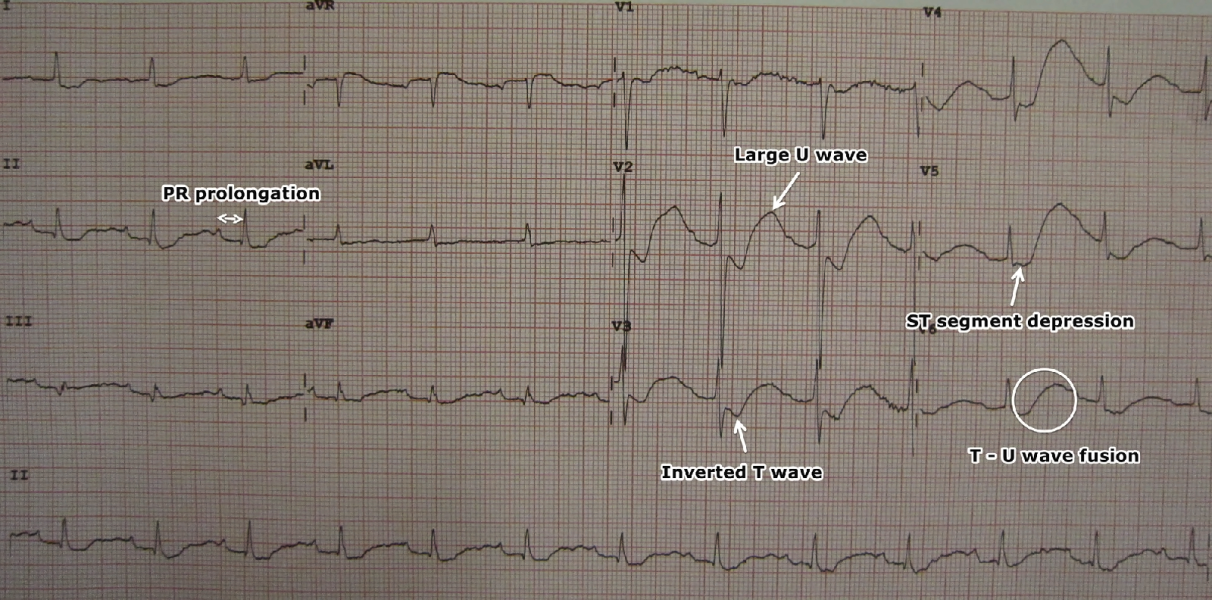
|[[image:LowKECG.png|thumb|700px|center|An ECG in a person with a potassium level of 1.1 showing the classical ECG changes of ST segment depression, inverted T waves, large U waves, and a slightly prolonged PR interval. By James Heilman, MD - Own work, CC BY-SA 3.0 | |[[image:LowKECG.png|thumb|700px|center|An ECG in a person with a potassium level of 1.1 showing the classical ECG changes of ST segment depression, inverted T waves, large U waves, and a slightly prolonged PR interval. By James Heilman, MD - Own work, CC BY-SA 3.0]] | ||
|} | |} | ||
<br style="clear:left" /> | <br style="clear:left" /> | ||
[[image:LowKECG.png|thumb|700px|right|An ECG in a person with a potassium level of 1.1 showing the classical ECG changes of ST segment depression, inverted T waves, large U waves, and a slightly prolonged PR interval. By James Heilman, MD - Own work, CC BY-SA 3.0]] | |||
<br style="clear:left" /> | |||
{{#ev:youtube|7TWu0_Gklzo}} | |||
==Table== | |||
{| | |||
! align="center" style="background:#4479BA; color: #FFFFFF;" + |Complications | |||
! align="center" style="background:#4479BA; color: #FFFFFF;" + |Polymyositis | |||
! align="center" style="background:#4479BA; color: #FFFFFF;" + |Dermatomyositis | |||
|- | |||
! align="center" style="background:#DCDCDC;" + |[[Cancer|Malignancy]] | |||
| align="left" style="background:#F5F5F5;" + | | |||
*[[Lung]] | |||
| align="center" style="background:#F5F5F5;" + | | |||
*[[Lung]] | |||
|} | |||
Latest revision as of 02:27, 23 May 2019
pic

{{#ev:youtube|7TWu0_Gklzo}}
Table
| Complications | Polymyositis | Dermatomyositis |
|---|---|---|
| Malignancy |