Uploads by NNikravangolsefid
Jump to navigation
Jump to search
This special page shows all uploaded files.
| Date | Name | Thumbnail | Size | Description | Versions |
|---|---|---|---|---|---|
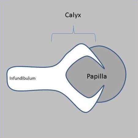
| 06:03, 21 August 2020 | Lobster-claw-sign-papillary-necrosis.jpg (file) |  |
12 KB | Case courtesy of Dr Matt A. Morgan, <a href="https://radiopaedia.org/">Radiopaedia.org</a>. From the case <a href="https://radiopaedia.org/cases/40421">rID: 40421</a> | 1 |
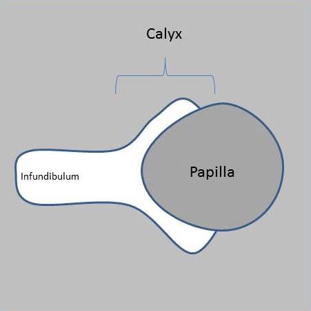
| 06:02, 21 August 2020 | Lobster-claw-sign-papillary-necrosis-normal.jpg (file) |  |
11 KB | Case courtesy of Dr Matt A. Morgan, <a href="https://radiopaedia.org/">Radiopaedia.org</a>. From the case <a href="https://radiopaedia.org/cases/40421">rID: 40421</a> | 1 |
| 08:27, 27 July 2020 | Blausen 0592 KidneyAnatomy 01.png (file) |  |
1.17 MB | 1 | |
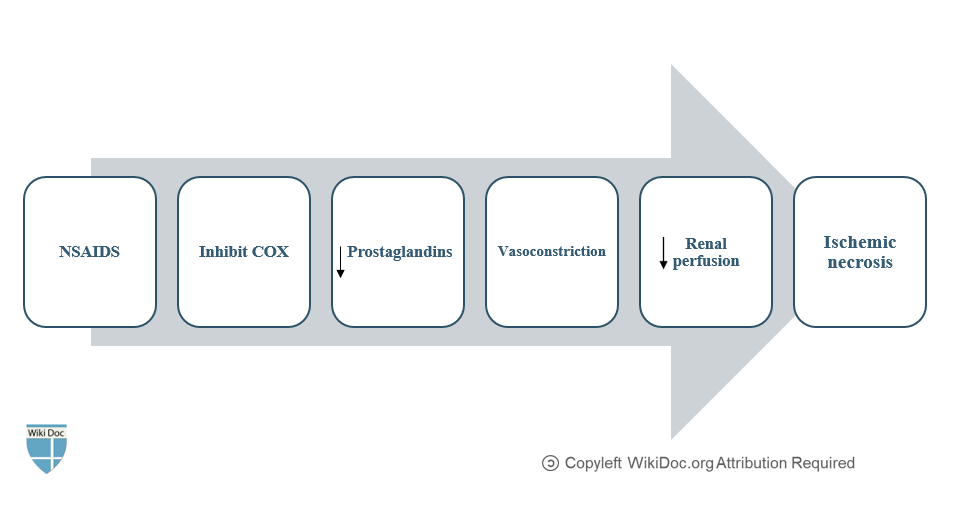
| 08:09, 27 July 2020 | Papillary necrosis-NSAIDS mechanism.PNG (file) |  |
26 KB | 1 | |
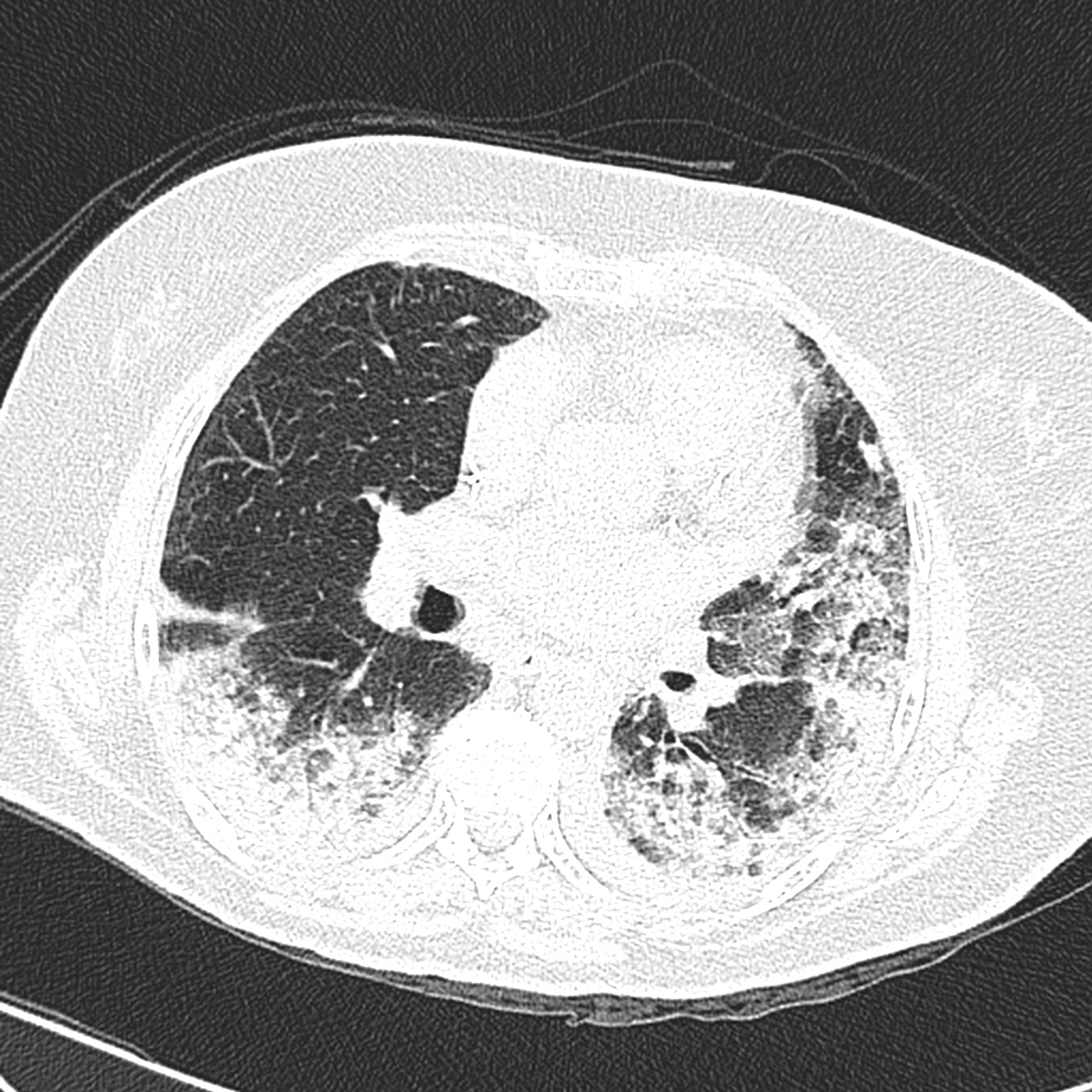
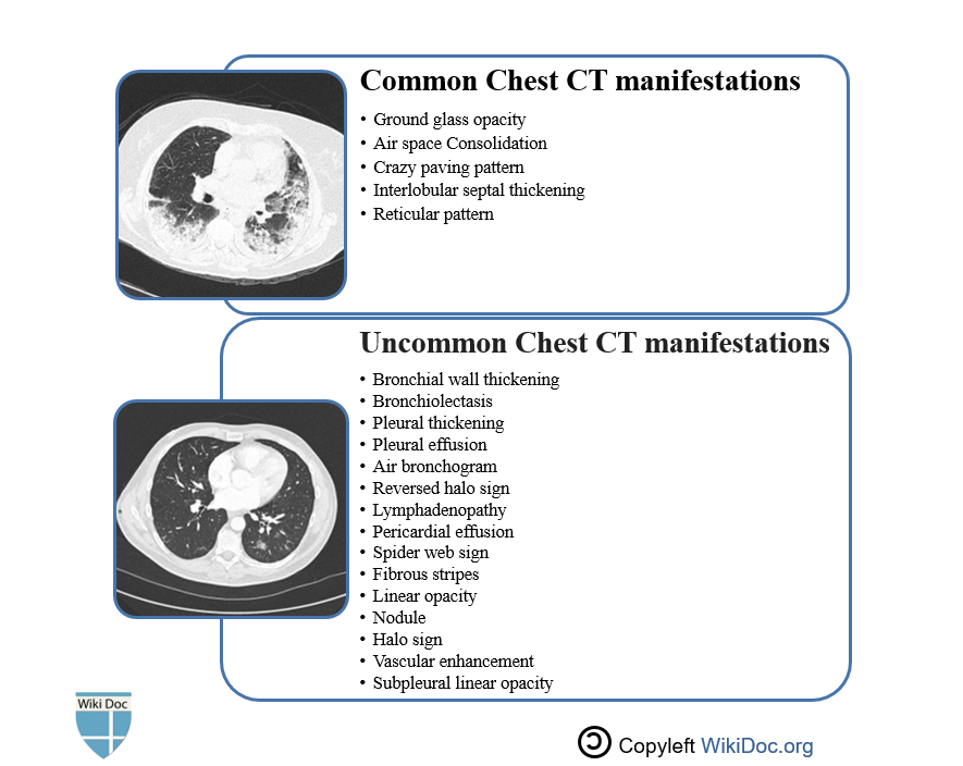
| 19:00, 19 July 2020 | CT-FINDINGS-COVID-19.PNG (file) |  |
158 KB | 1 | |
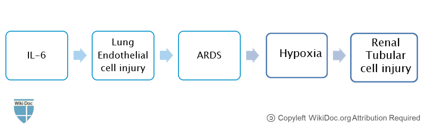
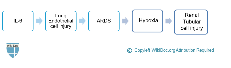
| 18:56, 19 July 2020 | INTERLEUKIN-6-AKI.PNG (file) |  |
22 KB | 1 | |
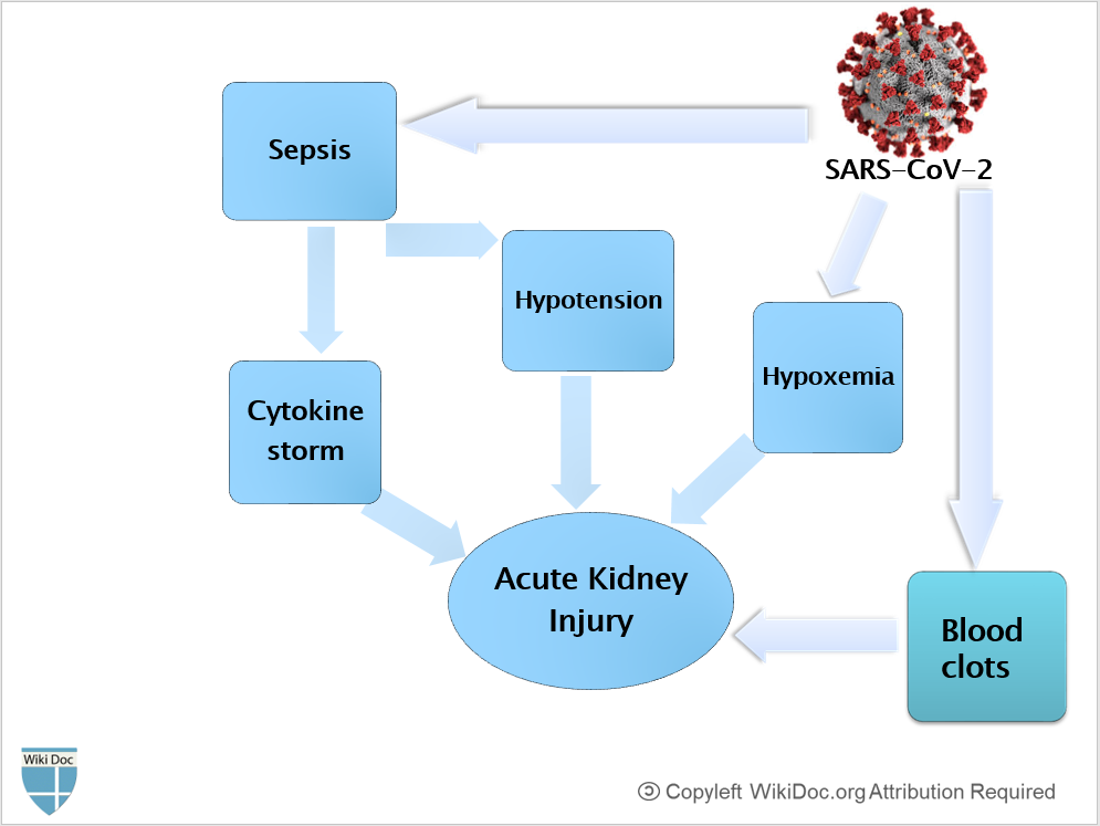
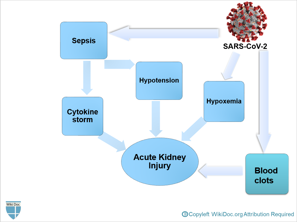
| 18:53, 19 July 2020 | AKI PATHOPHYSIOLOGY-COVID19.PNG (file) |  |
148 KB | 1 | |
| 18:49, 19 July 2020 | AKI physiopathology COVID19.PNG (file) |  |
148 KB | 2 | |
| 08:59, 19 July 2020 | CT-findings-covid19.pptx (file) | 311 KB | CT manifestations of COVID-19 | 1 | |
| 08:56, 19 July 2020 | AKI physiopathology-covid19.pptx (file) | 168 KB | 1 | ||
| 08:51, 19 July 2020 | CT-COVID-19.PNG (file) |  |
161 KB | 1 | |
| 06:36, 19 July 2020 | INTERLEUKIN6-AKI.PNG (file) |  |
28 KB | 1 | |
| 06:33, 19 July 2020 | IL-6-AKI-COVID-19.PNG (file) |  |
28 KB | 2 | |
| 06:27, 19 July 2020 | AKI physiopathology COVID.PNG (file) |  |
150 KB | 5 | |
| 21:26, 6 July 2020 | Covid-19-pneumonia-26.jpg (file) |  |
80 KB | Bilateral multilobar peripheral ground-glass opacities in both lungs predominantly in mid to lower zones. | 1 |
| 21:06, 6 July 2020 | Covid-19-pneumonia-122.jpg (file) |  |
893 KB | multilobar large areas of ground-glass opacities with septal thickening and a crazy-paving pattern | 1 |
| 11:58, 28 June 2020 | CT-manifestations-COVID19.PNG (file) |  |
153 KB | 1 | |
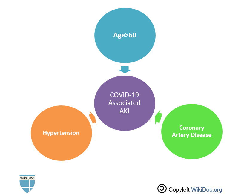
| 11:45, 28 June 2020 | COVID-19 associated AKI.PNG (file) |  |
33 KB | 1 | |
| 23:52, 19 June 2020 | Histopathology of secondary segmental glomerular sclerosis of hypertensive nephropathy.jpg (file) |  |
128 KB | Light micrograph of glomerulus showing secondary segmental sclerosis of hypertensive nephropathy | 1 |
| 22:51, 7 June 2020 | Renal arterial hyalinosis - pas - very high mag.jpg (file) |  |
3.91 MB | Renal arterial hyalinosis | 1 |
| 21:34, 5 June 2020 | Histopathology of hypertensive glomerular lesion of hypertensive nephropathy.jpg (file) |  |
114 KB | Light micrograph showing hypertensive glomerular lesion of hypertensive nephropathy: global glomerular collapse and filling of Bowman’s space with a lightly staining collagenous material | 1 |
| 20:37, 5 June 2020 | Fibrous intimal thickening in hypertensive nephropathy.jpg (file) |  |
41 KB | 1 | |
| 03:57, 20 May 2020 | Nasrinnik.jpg (file) |  |
2.1 MB | profile picture | 1 |