Skin cancer physical examination
Jump to navigation
Jump to search
|
Skin cancer Microchapters |
Editor-In-Chief: C. Michael Gibson, M.S., M.D. [1]
Overview
There are a variety of different skin cancer symptoms. These include crabs or changes in the skin that do not heal, ulcers in the skin, discoloration, and changes in existing moles.
Physical Examination
- Basal cell carcinoma usually looks like a raised, smooth, pearly bump on the sun-exposed skin of the head, neck or shoulders. Sometimes small blood vessels can be seen within the tumor. Crusting and bleeding in the center of the tumor frequently develops. It is often mistaken for a sore that does not heal.
- Squamous cell carcinoma is commonly a red, scaling, thickened patch on sun-exposed skin. Ulceration and bleeding may occur. When SCC is not treated, it may develop into a large mass.
- Most melanomas are brown to black looking lesions. Signs that might indicate a malignant melanoma include change in size, shape, color or elevation of a mole. The appearance of a new mole during adulthood, or new pain, itching, ulceration or bleeding of an existing mole should be checked.
The following image is an example of how skin cancer can affect the nails.
-
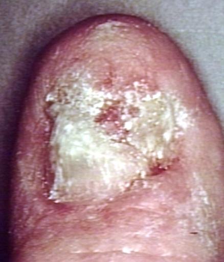 Skin carcinoma
