Non-Hodgkin lymphoma chest x ray
Jump to navigation
Jump to search
|
Non-Hodgkin lymphoma Microchapters |
|
Differentiating Non-Hodgkin's Lymphoma |
|---|
|
Treatment |
|
Case Studies |
|
Non-Hodgkin lymphoma chest x ray On the Web |
|
American Roentgen Ray Society Images of Non-Hodgkin lymphoma chest x ray |
|
Risk calculators and risk factors for Non-Hodgkin lymphoma chest x ray |
Editor-In-Chief: C. Michael Gibson, M.S., M.D. [1]; Associate Editor(s)-in-Chief: Preeti Singh, M.B.B.S.[2]
Overview
On chest x ray, non-Hodgkin lymphoma is characterized by nodules and pleural effusion.
Chest X ray
On chest x ray, non-Hodgkin lymphoma is characterized by:[1]
- Presence of nodules and hilar masses suggestive of central lymphadenopathy
- Pericardial effusions
- Pleural effusion
- Parenchymal involvement may be seen
- The mediastinal mass ratio can be determined by chest X-ray.
- A mediastinal mass ratio of greater than 0.33 is consistent with bulky disease.
-
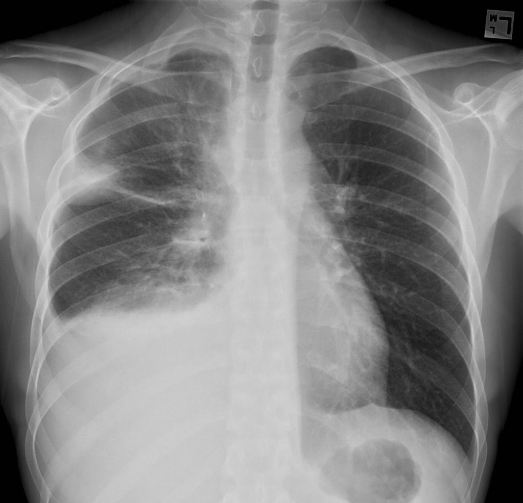 Large unilateral right side pleural effusion can be seen with no fluid on the left. There is no evidence of cardiomegaly. There is an lesion with increased opacity on the peripheral of right upper lung field and another lesion located at the right hilum. Image courtesy of Dr. Jack Ren. Radiopaedia

References
- ↑ Bligh MP, Borgaonkar JN, Burrell SC, MacDonald DA, Manos D (2017). "Spectrum of CT Findings in Thoracic Extranodal Non-Hodgkin Lymphoma". Radiographics. 37 (2): 439–461. doi:10.1148/rg.2017160077. PMID 28287948.