Isosporiasis laboratory findings
|
Isosporiasis Microchapters |
|
Diagnosis |
|---|
|
Treatment |
|
Case Studies |
|
Isosporiasis laboratory findings On the Web |
|
American Roentgen Ray Society Images of Isosporiasis laboratory findings |
|
Risk calculators and risk factors for Isosporiasis laboratory findings |
Editor-In-Chief: C. Michael Gibson, M.S., M.D. [1]
Laboratory Findings
Microscopic demonstration of the large, typically shaped oocysts, is the basis for diagnosis. Because the oocysts may be passed in small amounts and intermittently, repeated stool examinations and concentration procedures are recommended. If stool examinations are negative, examination of duodenal specimens by biopsy or string test (Enterotest®) may be needed. The oocysts can be visualized on wet mounts by microscopy with bright-field, differential interference contrast (DIC), and UV fluorescence. They can also be stained by modified acid-fast stain.

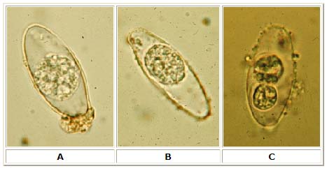
A, B, C: Oocysts of Isospora belli. The oocysts are large (25 to 30 µm) and have a typical ellipsoidal shape. When excreted, they are immature and contain one sporoblast (A, B). The oocyst matures after excretion: the single sporoblast divides in two sporoblasts (C), which develop cyst walls, becoming sporocysts, which eventually contain four sporozoites each. Images contributed by Georgia Division of Public Health.

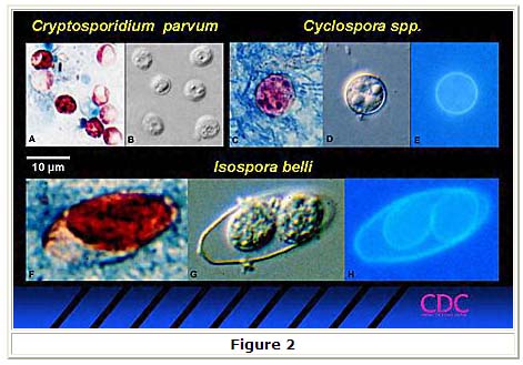
Oocysts of Isospora belli can also be stained with acid-fast stains, and can be visualized by UV fluorescence on wet mounts, as illustrated in Figure 2. Three coccidian parasites that most commonly infect humans, seen in acid-fast stained smears (2A, 2C, 2F), bright-field differential interference contrast (2B, 2D, 2G) and UV fluorescence (2E, 2H, C. parvum oocysts do not autofluoresce).