Gallbladder polyp ultrasound
Jump to navigation
Jump to search
|
Gallbladder Polyp Microchapters |
|
Diagnosis |
|---|
|
Treatment |
|
Case Studies |
|
Gallbladder polyp ultrasound On the Web |
|
American Roentgen Ray Society Images of Gallbladder polyp ultrasound |
|
Risk calculators and risk factors for Gallbladder polyp ultrasound |
Editor-In-Chief: C. Michael Gibson, M.S., M.D. [1]
Please help WikiDoc by adding more content here. It's easy! Click here to learn about editing.
Overview
Ultrasound
- Gallbladder polyps are typically identified on ultrasonography, which has a sensitivity and specificity of over 90%.
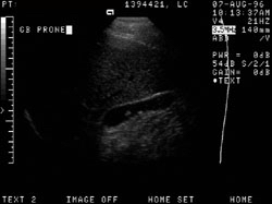
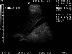
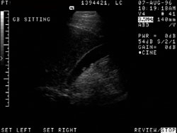
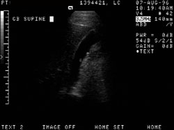
Shown below are ultrasound images of gallbladder polyps. Images courtesy of Professor Peter Anderson DVM PhD and published with permission © PEIR, University of Alabama at Birmingham, Department of Pathology
-
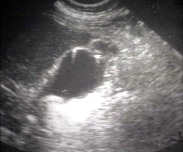 USG: Gallbladder: Adenomatous polyp
 -
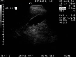
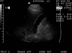 51-year-old male with right lower quadrant pain. Two small polyps are seen within the gallbladder (the largest of which measures 7mm)
 -
 Two small polyps are seen within the gallbladder (the largest of which measures 7mm).


-
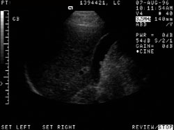
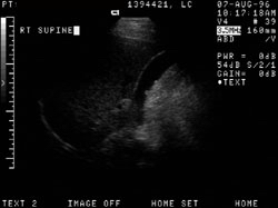
 Two small polyps are seen within the gallbladder (the largest of which measures 7mm)
 -
 Two small polyps are seen within the gallbladder (the largest of which measures 7mm)
 -
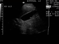
 Two small polyps are seen within the gallbladder (the largest of which measures 7mm)


-
 Two small polyps are seen within the gallbladder (the largest of which measures 7mm)
 -
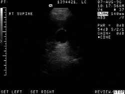 Two small polyps are seen within the gallbladder (the largest of which measures 7mm)
 -
 Two small polyps are seen within the gallbladder (the largest of which measures 7mm)


-
 Two small polyps are seen within the gallbladder (the largest of which measures 7mm)
 -
 Two small polyps are seen within the gallbladder (the largest of which measures 7mm)


- ERCP (Endoscopic Retrograde Cholangiopancreatography)
- Contrast enhanced CT may aid in the diagnosis with an overall accuracy of 87% for cancer.
- HIDA Scan (Cholescintigraphy): Hepatobiliary iminodiacetic acid scan
- FDG-PET adds little to the CT.
- Importantly, endoscopic ultrasound, which permits detailed evaluation of the gallbladder wall, has excellent diagnostic capabilities and should be used for indeterminate polyps of 5-15 mm.