Epicardial tumor pathophysiology
Jump to navigation
Jump to search
|
Epicardial tumor Microchapters |
|
Diagnosis |
|---|
|
Treatment |
|
Case Studies |
|
Epicardial tumor pathophysiology On the Web |
|
American Roentgen Ray Society Images of Epicardial tumor pathophysiology |
|
Risk calculators and risk factors for Epicardial tumor pathophysiology |
Editor-In-Chief: C. Michael Gibson, M.S., M.D. [1]
Please help WikiDoc by adding more content here. It's easy! Click here to learn about editing.
Overview
Pathophysiology
Microscopic pathology
-
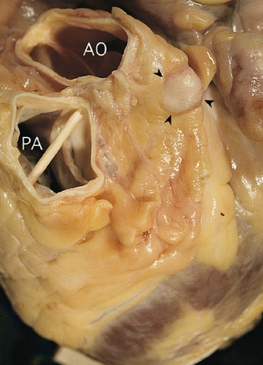 Granular cell tumor: Localized epicardial tumor (arrowheads) overlying the left main coronary artery close to its takeoff. Note aorta (AO) posterior to the pulmonary artery (PA).
 -
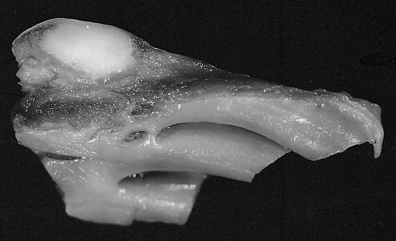 Granular cell tumor: Localized epicardial tumor: Note the cut surface of circumscribed white tumor on the epicardial surface and the underlying right ventricular trabeculations.

