Diabetic neuropathy physical examination
|
Diabetic neuropathy Microchapters |
|
Diagnosis |
|---|
|
Treatment |
|
Case Studies |
|
Diabetic neuropathy physical examination On the Web |
|
American Roentgen Ray Society Images of Diabetic neuropathy physical examination |
|
Risk calculators and risk factors for Diabetic neuropathy physical examination |
Editor-In-Chief: C. Michael Gibson, M.S., M.D. [1]
Please help WikiDoc by adding content here. It's easy! Click here to learn about editing.
Overview
Physical Examination
A study by the International Cooperative Group for Clinical Examination Research found that the monofilament and reflex testing were the most reproducible[1]. This study also found that monofilament testing of four locations per foot was adequate.
A systematic review by the Rational Clinical Examination found that "Abnormal results on monofilament testing and vibratory perception (alone or in combination with the appearance of the feet, ulceration, and ankle reflexes) are the most helpful signs"[2].
Images
(Images courtesy of Charlie Goldberg, M.D., UCSD School of Medicine and VA Medical Center, San Diego, California)
-
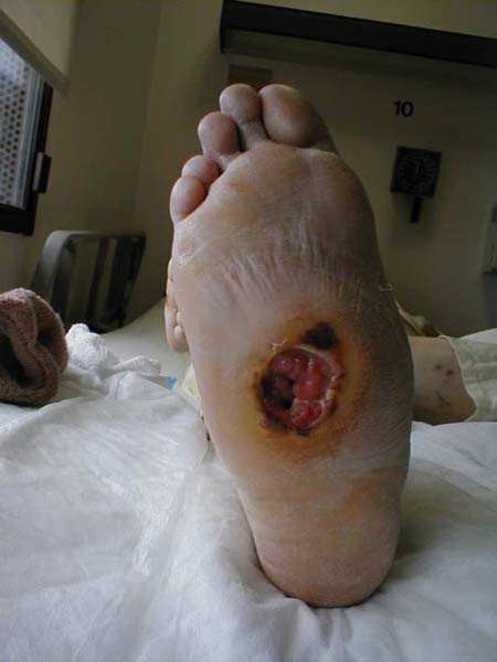
 Diabetes induced neuropathy has lead to large, painless ulcer on bottom of foot. Patient has had toes removed several years earlier due to prior infection.

-
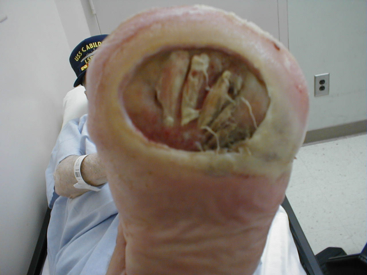
 Severe diabetes induced neuropathy has resulted in Charcot foot deformity. This ultimately lead to large painless ulcer on bottom of foot.
 -
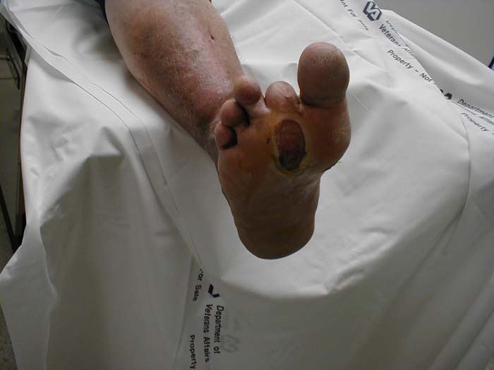 Diabetes induced neuropathy that has lead to painless ulcer on bottom of foot.
 -
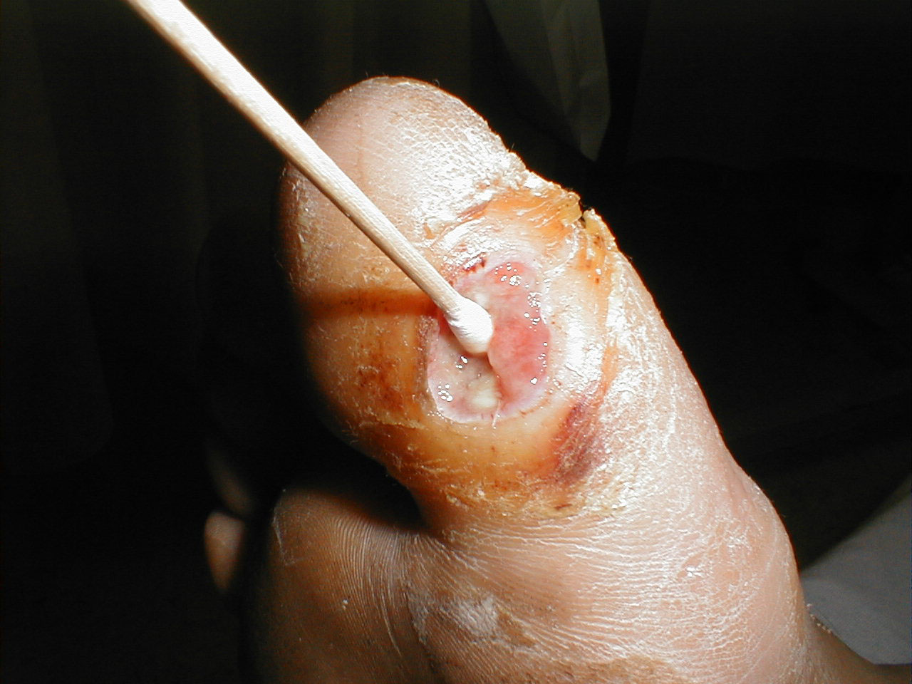 Diabetes induced neuropathy led to development of the ulcer shown above. The Q-tip easily passes to the level of the underlying bone, clinical evidence of osteomyelitis. Incidentally, this is not painful to the patient as he is insensate.


References
- ↑ Smieja M, Hunt DL, Edelman D, Etchells E, Cornuz J, Simel DL (1999). "Clinical examination for the detection of protective sensation in the feet of diabetic patients. International Cooperative Group for Clinical Examination Research". J Gen Intern Med. 14 (7): 418–24. doi:10.1046/j.1525-1497.1999.05208.x. PMC 1496604. PMID 10417599.
- ↑ Kanji JN, Anglin RE, Hunt DL, Panju A (2010). "Does this patient with diabetes have large-fiber peripheral neuropathy?". JAMA. 303 (15): 1526–32. doi:10.1001/jama.2010.428. PMID 20407062. Review in: Ann Intern Med. 2010 Oct 19;153(8):JC4-10