Anterior horn of the lateral ventricle
Template:Infobox Brain Editor-In-Chief: C. Michael Gibson, M.S., M.D. [1]
The ventricular system is a set of structures in the brain continuous with the central canal of the spinal cord.
Components
The system comprises four ventricles:
- right and left lateral ventricles
- third ventricle
- fourth ventricle
there are a few little holes in the brain leading from these ventricles, though only the first two of the list below are generally considered part of the ventricular system:
| Name | From | To |
| right and left interventricular foramina (Monro) | lateral ventricles | third ventricle |
| cerebral aqueduct (Sylvius) | third ventricle | fourth ventricle |
| Median aperture (Magendie) | fourth ventricle | subarachnoid space/cisterna magna |
| right and left Lateral aperture (Luschka) | fourth ventricle | subarachnoid space/cistern of great cerebral vein |
Each ventricle contains a choroid plexus that produces cerebrospinal fluid (CSF) used to bathe and cushion the brain and spinal cord within their bony confines.
Ventricles
There are four cerebral ventricles: the paired lateral ventricles, and midline the third and fourth ventricles. The two lateral ventricles, located within the cerebrum, are relatively large and C-shaped, roughly wrapping around the dorsal aspects of the basal ganglia. It is in the lateral ventricles of the embryo that the successive generation of neurons gives rise to the 6-layered structure of the neocortex, constructed from the inside out during development. Each lateral ventricle extends into the frontal, temporal and occipital lobes via the frontal (anterior), temporal (inferior), and occipital (posterior) horns, respectively.
The lateral ventricles both communicate via the interventricular foramina with the third ventricle, found centrally within the diencephalon. The third ventricle communicates via the cerebral aqueduct, located within the midbrain, with the fourth ventricle, found within the hindbrain. The three foramina to the subarachnoid space are found here, permitting cerebrospinal fluid produced in the ventricles to surround the brainstem, cerebellum, and cerebral cortex. The fourth ventricle is also continuous with the central canal, allowing CSF to bathe the inside surface of the spinal cord as well.
Flow of cerebrospinal fluid
Cerebrospinal fluid is produced by modified ependymal cells of the choroid plexus found in all components of the ventricular system except for the cerebral aqueduct and the occipital and frontal horns of the lateral ventricles. CSF flows from the lateral ventricles via the foramina of Monro into the third ventricle, and then the fourth ventricle via the cerebral aqueduct in the brainstem. From there it can pass into the central canal of the spinal cord or into the cisterns of the subarachnoid space via three small foramina: the central foramen of Magendie and the two lateral foramina of Luschka.
The fluid then flows around the superior sagittal sinus to be reabsorbed via the arachnoid villi into the venous system. CSF within the spinal cord can flow all the way down to the lumbar cistern at the end of the cord around the cauda equina where lumbar punctures are performed.
The aqueduct between the third and fourth ventricles is very small, as are the foramina, which means that they can be easily blocked, causing high pressure in the lateral ventricles. This is a common cause of hydrocephalus--otherwise known as water in the brain--and is an extremely serious condition due to both the damage caused by the pressure as well as nature of whatever caused the block (possibly a tumour or inflammatory swelling).
Protection of the brain
The brain and spinal cord are covered by a series of tough membranes called meninges, which protect these organs from rubbing against the bones of the skull and spine. The cerebrospinal fluid within the skull and spine is found between the pia mater and the arachnoid meninges and provides further cushioning.
Role in disease
Diseases of the ventricular system include abnormal enlargement (hydrocephalus) and inflammation of the CSF spaces (meningitis, ventriculitis) caused by infection or introduction of blood following trauma or hemorrhage.
Interestingly, scientific study of CAT scans of the ventricles in the late 1970s revolutionized the study of mental illness. Researchers found that patients with schizophrenia had enlarged ventricles compared to healthy subjects. This became the first "evidence" that mental illness was biological in origin and led to a reinvigoration of the study of such conditions via modern scientific techniques. Whether the enlargement of the ventricles is a cause or a result of schizophrenia has not yet been ascertained, however. Nowadays, magnetic resonance imaging (MRI) has superseded the use of CAT in research into the role of ventricular abnormalities in psychiatric illness.
Development
The structures of the ventricular system are embryologically derived from the centre of the neural tube (the neural canal).
In brainstem
elaini is a primitive neural tube that will become the brain stem develops, the neural canal expandsleft and right creating the fourth ventricle, whereas the neural canal that does not expand and remains the same at the level of the midbrain superior to the fourth ventricle forms the cerebral aqueduct. Likewise, the neural canal in the spinal cord that does not change forms the central canal.
Additional images
-
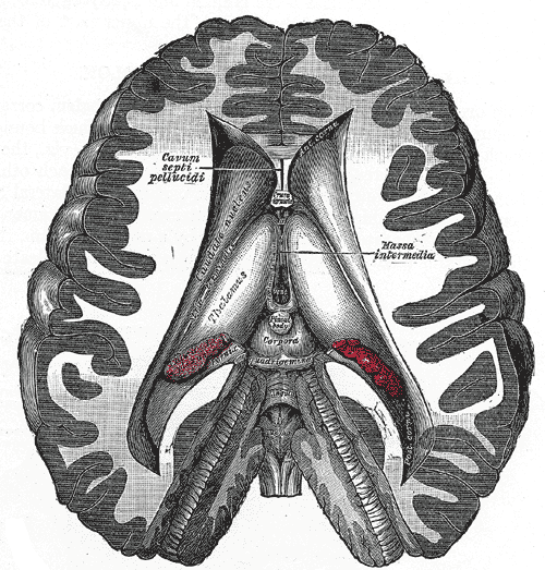 Coronal dissection showing the ventricles of the brain.

External links
- Template:BrainMaps
- ventricular system and CSF (concise description, Uni Washington)
- CSF at answers.com (brief description, excellent diagram of CSF flow)
References
- Purves D, Augustine GJ, Fitzpatrick D, Hall WC, Lamantia AS, McNamara JO, Williams SM, Neuroscience (third edition). Sinauer Associates Inc, July 2004. ISBN 0-87893-725-0
- Edgley S et al, Neuroanatomy from the Department of Anatomy, University of Cambridge.
Template:Ventricular system
de:Ventrikelsystem
he:חדרי המוח
nl:Ventrikelstelsel
no:Ventrikkelsystemet
fi:Aivokammio
sv:Ventrikelsystemet