2,3-Bisphosphoglycerate
Editor-In-Chief: C. Michael Gibson, M.S., M.D. [1]
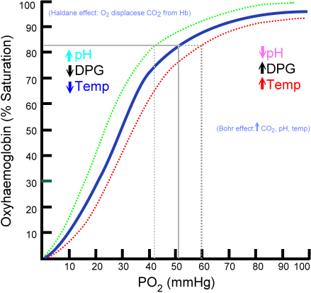
2,3-Bisphosphoglycerate (2,3-BPG, also known as 2,3-diphosphoglycerate or 2,3-DPG) is a three carbon isomer of the glycolytic intermediate 1,3-bisphosphoglycerate. 2,3-BPG is present in human red blood cells (RBC; erythrocyte) at approximately 5 mmol/L. It binds with greater affinity to deoxygenated hemoglobin (e.g. when the red cell is near respiring tissue) than it does to oxygenated hemoglobin (e.g. in the lungs). In bonding to partially deoxygenated hemoglobin it allosterically upregulates the release of the remaining oxygen molecules bound to the hemoglobin, thus enhancing the ability of RBCs to release oxygen near tissues that need it most.
Recently, scientists have found the similarities between low amounts of 2,3-BPG with the occurrence of HAPE at high altitudes.
Its function was discovered in 1967 by Reinhold Benesch and Ruth Benesch.[1]
Metabolism
2,3-BPG is formed from 1,3-BPG by an enzyme called bisphosphoglycerate mutase. It is broken down by a phosphatase to form 3-phosphoglycerate. Its synthesis and breakdown are therefore a way around a step of glycolysis.
                       glycolysis
              1,3-BPG ------------> 3-PG
                   \_               _^
                     \_           _/
              2,3-BGP  v         /  2,3-BPG
               mutase     2,3-BPG   phosphatase
Effects of binding

When 2,3-BPG binds deoxyhemoglobin, it acts to stabilize the low oxygen affinity state (T state) of the oxygen carrier. It fits neatly into the cavity of the deoxy- conformation, exploiting the molecular symmetry and positive polarity by forming salt bridges with lysine and histidine residues in the four subunits of hemoglobin. The R state, with oxygen bound to a heme group, has a different conformation and does not allow this interaction.
By selectively binding to deoxyhemoglobin, 2,3-BPG stabilizes the T state conformation, making it harder for oxygen to bind hemoglobin and more likely to be released to adjacent tissues. 2,3-BPG is part of a feedback loop that can help prevent tissue hypoxia in conditions where it is most likely to occur. Conditions of low tissue oxygen concentration such as high altitude (2,3-BPG levels are higher in those acclimated to high altitudes), airway obstruction, or congestive heart failure will tend to cause RBCs to generate more 2,3-BPG in their effort to generate energy by allowing more oxygen to be released in tissues deprived of oxygen. Ultimately, this mechanism increases oxygen release from RBCs under circumstances where it is needed most. This release is potentiated by the Bohr effect in tissues with high energetic demands.
Fetal hemoglobin
Interestingly, fetal hemoglobin (HbF) exhibits a low affinity for 2,3-BPG, resulting in a higher binding affinity for oxygen. This increased oxygen binding affinity relative to that of adult hemoglobin (HbA) is due to HbF having two α/γ dimers as opposed to the two α/β dimers of HbA. The positive histidine residues of HbA β-subunits that are essential for forming the 2,3-BPG binding pocket are replaced by serine residues in HbF γ-subunits.
References
- ↑ Benesch, R. (1967). "The effect of organic phosphates from the human erythrocyte on the allosteric properties of hemoglobin". Biochem Biophys Res Commun. 26 (2): 162–7. Unknown parameter
|coauthors=ignored (help)
- Berg, J.M., Tymockzko, J.L. and Stryer L. Biochemistry (5th ed). W.H. Freeman and Co, New York, 1995. ISBN 0-7167-4684-0.
- Template:GPnotebook
- Online medical dictionary